-->

TEST

Monday 30 April 2012

Mini cargo transporters on a rat run

Molecular motors are the key to the development of higher forms of life. They transport proteins, signal molecules and even entire chromosomes down long protein fibers, components of the so-called cytoskeleton, from one location in the cell to another. Not unlike trucks on a motorway, there are permanently thousands of these small motor proteins underway at any given point in time – a highly coordinated and extremely fast mode of transport. This highly efficient infrastructure is a prerequisite for the formation of large, complex cells and multicellular organisms. Bacteria, for example, lack this foundation, because they possess neither molecular motors nor cytoskeletons.
Biophysicists of the Technische Universitaet Muenchen and the Ludwig Maximillians Universitaet Muenchen have discovered why some of these transporters can, like cars on a multi-lane motorway, change lanes: The heads of one kinesin (red) have a longer range than the other (blue) which allows "lane change" between the individual fibers (protofilaments) of the microtubule and results in a spiraling movement of the motor on the microtubule. A shorter range of the heads results in a straight movement of the motor. Credit: Melanie Brunnbauer /TU Muenche
Kinesins represent one class of such molecular motors. They run along microtubules comprising 13 individual fibers arranged in a tube form. Kinesins are made up of a twisted pair of protein chains. Each chain comprises a head that can dock to the surface of the microtubules and a neck domain, as well as a stalk and tail domain that the cargo is attached to. Kinesins move forward by placing one head in front of the other in alternation which resembles human walking. The first mechanistically scrutinized kinesin was Kinesin-1, which performs numerous steps in succession without detaching from the microtubule. In the process it moves ahead in a perfectly straight path on its long journey, always remaining on a single fiber of the microtubule.


 kinesin type-1 straight movement

Scientists led by Zeynep Oekten, group leader at the Biophysics Department of the Technische Universitaet Muenchen, and Melanie Brunnbauer, a doctoral candidate at the Biophysics Department, have now for the first time demonstrated that kinesins also "switch lanes" during transport. The scientists identified the region in the kinesin protein that determines whether a given kinesin type moves on a straight path or in a spiral fashion. It is a structural element in the neck domain. "If the neck region is stable, the two kinesin heads have only limited reach. The kinesin cannot make any sidesteps and thus moves straight ahead," says Oekten. "However, if the responsible area becomes destabilized, the reach of the heads is increased and the motor protein can jump fibers and spiral around the microtubule."
To confirm this new insight, the scientists integrated specific amino acids into the responsible areas – a kind of molecular switch that allowed them to regulate the reach of the two heads. The result left no doubt: Destabilizing the neck region of the Kinesin-1 motor increases the reach of the two heads, which in turn causes the Kinesin-1 to depart from its normally perfectly straight path and move along a spiral-shaped path. When they mimicked a stable neck region using a chemical crosslinker, they coerced the protein into running straight again.


 kinesin type-2 :lane change


Oekten and Brunnbauer arrived at their new insight using a unique experimental setup. They placed two 3-micron large synthetic beads in a solution and trapped each using a laser beam, a so-called pair of "optical tweezers." Then, in precision work, they placed a piece microtubule between the beads. In a final step, again using a laser beam, they trapped a third bead coated with a specific type of kinesin and carefully placed it onto the microtubule.
As soon as they deactivated the third laser beam, the motor protein started marching forward and the scientist could follow the path of the molecule under the microscope. "In this way we were able, for the first time ever, to directly observe the spiraling movement of a motor type," explains Oekten. "When we saw the teetering movement of a Kinesin-2 protein for the first time, we all laughed. The motion was so clear and obvious, you just had to look at it and all doubt vanished." The experimental setup allows the molecular motors to move freely, thereby emulating real-life conditions in the cell much better than previous methods of investigation.
     
                                            molecular dynamic simulation of kinesin step


Using their new experimental setup, Oekten and Brunnbauer investigated a whole series of different Kinesin-2 proteins from various organisms – with an unexpected result: Contrary to the hitherto prevalent assumption that kinesins typically move only on straight paths, almost all kinesins displayed some form of spiral movement, in manifold variations. "This shows us that spiral motion is not an exception in nature, but rather the rule," explains Oekten. "In fact, the more relevant question is why evolution has brought about the straight-line movement as we observe with the Kinesin-1. That is truly unusual considering the nano-scale precision it requires to confine a kinesin transporter on an exclusively straight path." The researchers Oekten and Brunnenbauer hope to more closely investigate the reasons for the various kinds of motion in the future.


read more

Thursday 26 April 2012

DST JRF Vacancy in Biology at IISER, Thiruvananthapuram

Applications are invited from Indian nationals for the position of “JRF” in a DST sponsored project entitled “To determine the molecular mechanisms involved in centrosomal Transforming Acidic Coiled-Coil 3 (TACC3) mediated cell cycle progression”

No. of positions:  1 Qualifications:Master’s degree in any area of Biological Sciences with minimum of 55% marks, qualified CSIR-UGC NET-JRF/LS, or GATE (valid qualified score).

Experience: Applicants should have experience in molecular biology techniques including gene cloning; cell biology techniques including mammalian tissue culture, genetic manipulation in mammalian cells. Prior experience of working in research projects involving molecular cloning, mammalian tissue culture, genetic manipulation, generation of stable cell lines is desirable.

Age limit:28 yrs or below. A relaxation of 3-5 yrs will be applicable to candidates belonging to SC/ST/OBC, Persons with Disability and women category.Fellowship: UGC-NET JRF: monthly 16000/- + HRA; NET-LS or GATE: 12000/- + HRA.Duration:Initial appointment for one year, extendable up to 3 yrs based on performance.

How to apply:Application should contain a detail resume, contact details including phone number, email and postal address, a photograph (pasted on the resume), photocopies of educational/professional qualifications, reprints of papers etc. Research experience should be supported by certificate from previous employer.  Candidates should bring originals of the certificate for the qualifying degrees, age and National Examinations as well as the category certificate, if applicable, during the interview. Applications failing to meet minimum criteria will not be considered. Completed applications should reach Dr. Tapas Manna, School of Biology, Principal Investigator, IISER-Thiruvananthapuram, CET campus, Engineering College P.O. Thiruvananthapuram- 695016, Kerala by May 28, 2012. No TA/DA will be paid for appearing for the interview.

for further info visit :http://iisertvm.ac.in/appointments/projects/24/applications-invited-for-jrf-position-in-biology.html
read more

Applications are invited for temporary posts for the DBT/ ICAR funded projects.

Applications are invited for the following purely temporary posts for the DBT/ ICAR funded
projects.

1. Research Associate (One): Ph.D. with atleast three years research experience in
molecular biology after completion of Ph.D.

2. Technical Assistant (One): Graduate with ten years experience in handling the
Sequencer/ Computer Servers/ Databases/ Accounts.

3. Lab Assistant (One): Intermediate (10+2) with seven years of experience in field data
collection and in maintenance of laboratory.

4. SRF (Two): Masters degree in Life Sciences with at least 55% marks.

Application on plain paper with full bio-data and attested copies of mark sheets and certificates
should reach the undersigned before 15th May  of the publication of this advertisement on the
website of University of Delhi. The interview for eligible candidates will be conducted in the
Library, Department of Zoology, University of Delhi. Candidates will be intimated about the
interview date and time. No TA/DA will be paid for attending the interview.

for further info visit :http://www.du.ac.in/fileadmin/DU/students/Pdf/du/career/2012/24412_Zoology_Advertisement.pdf

read more

In Protein Folding, Internal Friction May Play a More Significant Role Than Previously Thought

Protein folding is the process by which not-yet folded chains of amino acids assume their specific shapes, hence taking on their specific functions. These functions vary widely: In the human body, proteins fold to become muscles, hormones, enzymes, and various other components.

"This protein folding process is still a big mystery," said UC Santa Barbara physicist Everett Lipman, one of several authors of a paper, "Quantifying internal friction in unfolded and intrinsically disordered proteins with single-molecule spectroscopy." The paper was published in the Proceedings of the National Academy of Sciences.

A protein's final shape, said Lipman, is primarily determined by the sequence of amino acid components in the unfolded chain. In the process, the components bump up against each other, and when the right configuration is achieved, the chain passes through its "transition state" and snaps into place.
"What we would like to understand eventually is how the chemical sequence of a protein determines what it is going to become and how fast it is going to get there," Lipman said.
Using a microfluidic mixing technique pioneered in the UCSB physics department by former graduate student Shawn Pfeil, the research team, including collaborators from the University of Zurich and the University of Texas, was able to monitor extremely rapid reconfiguration of individual protein molecules as they folded.
In the microfluidic mixer, a "denaturant" chemical used to unravel the proteins was quickly diluted, enabling observation of folding under previously inaccessible natural conditions. The measurements demonstrated that internal friction plays a more significant role in the folding process than could be seen in prior experiments, especially when the protein starts in the more compact unfolded configuration it would have in a denaturant-free living cell.
"At those size scales, everything is dominated by friction," said Lipman, comparing the environment of a protein molecule in water to a human body in molasses. Friction between the molecule and its liquid environment is an issue, as well as the "dry" friction that is independent of the surrounding solvent.
Internal friction slows down the folding process by reducing the rate at which the amino acid chain explores different configurations that may lead to the transition state. The longer it takes to find its native state -- its final form -- the higher the likelihood it could get stuck in an unfolded state.
"When it is unfolded, it is more vulnerable to being trapped in a misfolded state, or to aggregation with other unfolded protein molecules," said Lipman. Aggregation of misfolded proteins is thought to be a contributor to many types of diseases, such as the amyloid plaques that are associated with Alzheimer's disease. Alternatively, the unfolded and not usable protein could be broken back up into its component amino acids by the cell.
While there is no confirmed link between internal friction and aggregation, or any pattern of friction for one protein that affects others in the same way, Lipman and his colleagues are getting closer to understanding the degree to which internal friction affects the protein folding process.
"These measurements show that under realistic conditions, internal friction plays a significant role in the dynamics of the unfolded state. If a model of the protein folding process doesn't account for this, it will need to be reconsidered," he said
read more

Monday 23 April 2012

Recruitment for the post of Research Associate I and Research Associate II in Department of Plant Sciences, School of Life Sciences, University of Hyderabad, Hyderabad

Applications are invited for the following post:

1. Post: Research Associate I
Qualification: Applicants should have experience in plant functional genomics, RNAi / VIGS, Next Gen Sequencing preferably in tomato or other crop plants and should be supported by publications. PhD in Biological Sciences is required.

2. Post: Research Associate II
Qualification: Applicants should have experience in proteomics-2D/DIGE/LCMS and also in characterizing PTMs and should be supported by publications. PhD in Biological Sciences is required.

Candidates interested in above positions should send their CV, a statement clearly explaining how their skills are relevant to the position and the name / contact information for three references. The candidates can send their application by email at rameshwar.sharma-at-gmail.com and/or syellamaraju-at-gmail.com on or before May 7th, 2012.
No TA/DA would be provided for attending the interview.

for further info visit  http://www.uohyd.ac.in/images/recruitment/tomanetpositions.pdf


read more

Recruitment for the post of Senior Research Fellow and Junior Research Fellow for DBT sponsored project in Department of Biochemistry, University of Hyderabad, Hyderabad

Applications are invited on a plain paper (along with copies of educational qualifications and experience) from eligible candidates for selection for positions of SRF and JRF under the two projects entitled “Sys TB: A Network Program for Resolving the Intracellular Dynamics of Host Pathogen Interaction in TB Infection” with specific objective ‘Tracking temporal modulation in proteome composition of the Mtb phagosome’ (BT/PR3260/BRB/10/967/2011)” and “‘Identification and characterization of new iron-dependent post-transcriptional regulome of Mycobacteria’ under Innovative Young Biotechnologist Award (IYBA), BT/05/IYBA/2011 sponsored by Department of Biotechnology (DBT) sanctioned to Dr. Sharmistha Banerjee, Department of Biochemistry, University of Hyderabad

Positions

1. Senior Research Fellow – 1 No.
2. Junior Research Fellow – 1 No.

One SRF for project BT/PR3260/BRB/10/967/2011 and one JRF for BT/05/IYBA/2011

1. Senior Research Fellow (SRF) @ Rs. 18,000/- + 30% HRA* pm (fixed) for UGC-CSIR-NET qualified appointees; Rs. 14,000 + HRA* pm for non NET qualified appointees

2. Junior Research Fellow (JRF) @ Rs. 16,000 + 30% HRA* pm (fixed) for UGC-CSIR-NET qualified appointees; Rs. 12,000 + HRA* pm for NET-lectureship qualified appointees

Qualifications

1. SRF: M.Sc. in the area of Biology with minimum two years of research experience. Working experience with Mycobacterial cultures and tissue/cell culture will be preferred. The work involves travelling and working in collaborator’s labs.

2. JRF: M.Sc. in any area of Biology/Chemistry. Candidates with NET-qualification would be preferred for JRF. Research experience and experience of handling animals, if any, would be preferred.

Duration Appointments are made for one year and can be extendible after review each year until the duration of the project
Only candidates who would fit into objectives of the projects would be called for the interview.

Applications (along with photocopies of their qualifications/experience and reprints of published work) may be sent to Dr. Sharmistha Banerjee, Reader, Department of Biochemistry, School of Life Sciences, University of Hyderabad, Hyderabad 500046 on or before 7th May 2012. The short-listed candidates would have to appear for an interview at the School of Life Sciences, Science Complex, University of Hyderabad, Near Gachibowli, Hyderabad 500 046 on the date intimated to them. No TA/DA would be paid for attending the interview.

for further info : http://www.uohyd.ac.in/images/recruitment/advertisement-manpower-biochem-sb.pdf
read more

Saturday 21 April 2012

Polymers perform non-DNA evolution


Scientists have found that six polymer alternatives to DNA can pass on genetic information, and have evolved one type to specifically bind target molecules.1 They say that their work reveals both broader chemical possibilities for these key life functions and provides a powerful tool for nanotechnology and medicine. 
HNA
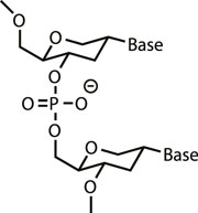
HNA could just as easily have been the molecule of life
 as DNA it now seems © Science/AAAS


'There is no overwhelming functional imperative for life to be based on DNA or RNA,' says Phil Holliger from the MRC Laboratory of Molecular Biology in Cambridge, UK, who led the team. 'Other polymers can perform these functions, at least at a basic level.' Holliger's team's xeno-nucleic acid (XNA) polymers each replace DNA's ribofuranose sugar ring with six other cyclic structures that can still form helical chains and base pairings. But rather than using relatively inefficient chemical synthesis, the scientists wanted to exploit polymerase and reverse transcriptase enzymes to copy genetic information from DNA templates to XNAs. In living organisms, polymerases can make RNA from nucleotide monomers using existing DNA strands as templates. Reverse transcriptases can then create a copy of the original DNA strand from that RNA in the same way.Yet those processes don't normally work with the kind of unnatural nucleotides the team used. Consequently, MRC scientist Vitor Pinheiro first mutated and then selected polymerase enzymes that best processed 1,5-anhydrohexitol nucleic acid (HNA) and cyclohexenyl nucleic acid (CeNA) nucleotide trisphosphates. As well as isolating an enzyme that would make long enough polymers with all six XNA types to encode genetic information, he similarly engineered reverse transcriptases. Together the enzymes could accurately replicate genetic information from DNA to XNA and back, but with enough copying mistakes for functions to evolve. 'For the best ones it's 99% accurate or better,' Holliger tells Chemistry World. 'You really don't need more than that.' 
XNA
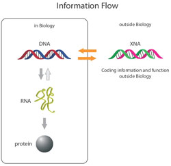
Xeno-nucleic acids can store genetic information that can be processed into
 DNA  and back again by mutated polymerases
© Science/AAAS


Repeated replication cycles were then used to evolve potent HNA aptamers, molecules that could act as drugs by recognising and binding to specific targets. In one example, starting from structures similar to DNA and RNA aptamers for HIV trans-activating response RNA (TAR), the team isolated HNAs that might be able to target HIV. These HNAs were then converted to DNA and back again, selecting for affinity to TAR over eight rounds of evolution, after which common genetic motifs emerged. 'Aptamers have the potential to rival antibodies in some clinical settings if we can get the properties right,' Holliger explains. 'This technology certainly has the potential to do that.'  Steven Benner, director of the Westheimer Institute  at  the Foundation for Applied Molecular Evolution in the US, notes that replacing DNA's sugar ring complements his team's introduction of non-standard nucleotide bases.2  'Together, these two classes of artificial chemical systems capable of heredity and evolution represents an expansion of our theories relating to the structure of molecules and the phenomenon of genetics,' he comments. 'It is relevant to biotechnology here on Earth as well as the possible forms that life might take throughout the cosmos.' 
Jack Szostak, who investigates processes that allowed early chemical and biological evolution on the Earth at Harvard University, US, calls Holliger's team's work 'very exciting'. 'This is very interesting with respect to the origin of life,' he says. 'In principle, many different polymers could serve the roles of RNA and DNA in living organisms. Why then does modern biology use only RNA and DNA? The answer probably lies in two "filters". First, only some nucleic acids could actually be made on the early Earth. Second, of those polymers that actually could be made, some may have been functionally superior to others in terms of ease or accuracy of replication, or ability to generate catalytic folded structures.'
read more

Tuesday 17 April 2012

Trial of Cancer-Fighting Virus Shows Promise

A new type of cancer treatment that uses a virus to infect and destroy tumor cells without harming normal cells is showing promise in early clinical trials.
The small, Phase 1 trial involved 23 patients with advanced cancers that had spread to multiple organs and who had exhausted other treatment options.
Each received an intravenous infusion of a virus called JX-594 at one of several dose levels. The virus was genetically engineered to contain an immune-stimulating gene to enhance its cancer-fighting properties, explained study co-senior author John Bell, a senior scientist at Ottawa Hospital Research Institution in Ontario, Canada.
Patients underwent biopsies eight to 10 days later. In seven of eight patients (87 percent) who received the highest two doses, researchers found evidence that the virus had not only infected the tumor cells while sparing healthy cells, but that the virus was replicating. Replication means that the virus is reproducing and infecting neighboring cancer cells, rather than just infecting tumor cells it directly came into contact with.
There was also evidence that the foreign immune-stimulating gene was expressed inside the tumor cells.
"This is a landmark observation in that it shows it's possible that a virus can find tumors, specifically grow in tumors but not in regular tissues, replicate and destroy them," Bell said.
The current trial was designed primarily to prove that it was both possible and safe to use a virus to infect tumor cells, and that the virus would then replicate. Side effects were minimal, with the main being brief and mild flu-like symptoms, researchers said.
Though larger trials are needed to determine efficacy, about 75 percent of patients in the two highest dose groups also saw a shrinking or stabilization of their tumor, while those in lower dose groups were less likely to experience this effect, according to the study.
"We didn't measure how well that specific immune-stimulating gene worked," Bell said. "But we definitely demonstrated the virus can go into the tumor, replicate only in the tumor and express a specific gene within the tumor."
The findings are published in the September issue of Nature.
One of the challenges in treating cancer is that cancer cells can spread into hard to find areas of the body, as well as in areas that can't be reached by a surgeon.
"The holy grail is a virus that could travel through the blood, find the tumors where they may be hiding, infect them and kill them," Bell explained.
Bell and his colleagues have been investigating cancer-fighting viruses for more than a decade. The virus, JX-594, is a distant relative of the smallpox virus; it's derived from a strain of vaccinia virus that has been used as a live vaccine in millions of people to vaccinate against small pox, Bell said.
In another bit of good news, the virus still infected the tumor cells even though all of the people in the study had previously been exposed to it as part of their child vaccinations, according to the study.
People in the study had several types of inoperable, advanced cancers, including lung, colorectal, melanoma, thyroid, pancreatic and ovarian. The virus can infect any type of epithelial, or surface cell, which are found throughout the digestive, reproductive, respiratory and urinary systems.
Researchers are currently planning a larger randomized clinical trial for patients with liver cancer.
William Phelps, director of preclinical and translational cancer research for the American Cancer Society, characterized the research as "preliminary, but really exciting."
"Viruses have a great capacity for finding cells in certain parts of the body. They often tend to infect only certain types of cells," Phelps said. "If we can manipulate that and take advantage of the natural capacity of the virus to spread throughout the body and to very selectively infect only certain types of cells, that could be very promising."
In this case, the virus contains "payload," or an extra gene that stimulates the immune system. "When the virus expresses that gene, it causes the immune system to kill the cell," he added. "It's very clever."
read more

Thursday 12 April 2012

Recruitment at: Biocon

Biocon hiring Grads, Post Grads as Managers

Position:
  • Manager - Formulation Development
  • Executive/ Sr Executive
  • SR.Manager -Business Development and Analysis 
  • Brand Manager 
  • Senior Manager - Microbial Fermentation 
  • Sr. Scientific Manager - Molecular Biology 
  • Scientific Manager -Analytical Development 
Location: Bangalore

Eligibility:

Manager - Formulation Development: M.Pharm, PHD
Executive/Sr Executive: B.Sc, B.Tech, M.Sc
SR.Manager -Business Development and Analysis: B.Pharm, B.Sc, B.Tech, M.B.A
Brand Manager:  B.Pharm, B.Sc, M.B.A
Senior Manager - Microbial Fermentation: B.E, B.Tech, M.Tech
Sr. Scientific Manager - Molecular Biology:  PHD
Scientific Manager -Analytical Development: MScE, PHD


for further details visit Biocon candidate portal

read more

Monday 9 April 2012

New immune defense enzyme discovered

Neutrophil granulocytes comprise important defences for the immune system. When pathogenic bacteria penetrate the body, they are the first on the scene to mobilise other immune cells via signal molecules, thereby containing the risk. To this end, they release serine proteases – enzymes that cut up other proteins to activate signal molecules. Scientists at the Max Planck Institute of Neurobiology in Martinsried have now discovered a new serine protease: neutrophil serine protease 4, or NSP4. This enzyme could provide a new target for the treatment of diseases that involve an overactive immune system, such as rheumatoid arthritis

The functioning of the immune system is based on the complex interplay of the most diverse cells and mediators. For example, neutrophil granulocytes (a group of specialized white blood cells) react to bacteria by releasing substances called serine proteases. These enzymes are able to activate signal molecules, such as the chemokines, by cleaving them at a specific position on the molecule. The active signal molecules then guide other immune cells to the focus of inflammation in order to destroy the pathogens.
A research team led by Dieter Jenne at the Max Planck Institute of Neurobiology in Martinsried has come across a previously unknown protease in humans: neutrophil serine protease 4, or NSP4. "The special thing about this enzyme is that it cuts proteins that have the amino acid arginine at a particular point", says Dieter Jenne, research group leader at the Martinsried-based Institute. "This is where NSP4 differs from the other three known neutrophil serine proteases, which are similar in molecular structure, but have a different recognition motif." The scientists may be able to harness this difference to develop an active substance that specifically inhibits NSP4, thereby reducing the immune reaction.
However, serine protease activity comes at a cost. The enzymes not only heal inflammations, but sometimes cause them in the first place. If too many immune cells are activated, they can use their arsenal of aggressive chemical weapons against the body's own tissues. A number of chronic inflammatory diseases are based on precisely this effect. As a result, scientists are searching for substances that can block the neutrophil proteases. To date, however, none of the substances tested have been developed into effective drugs.
"So far, we don't know the identity of the NSP4 substrate, but we assume they must be signal molecules", says Dieter Jenne. Activated chemokines can recruit a vast number of neutrophils, and their sheer quantity alone is enough to cause tissue damage. "Proteases sometimes act as accelerants and can even trigger a chronic inflammation quite independently of bacterial intruders. If we dampened down the defences, we could counteract this effect", explains the scientist.
In terms of evolutionary history, NSP4 is the oldest of the four known neutrophil serine proteases. Using gene sequences, scientists have shown that the enzyme has hardly changed through hundreds of millions of years of evolution from bony fish to humans. "That would indicate that NSP4 regulates a fundamental process", says Dieter Jenne.
The fact that the enzyme remained undiscovered until now is because it occurs at a much lower concentration than the other three proteases. The Max Planck scientists came across it while searching the human genome for genes that encode serine proteases. In the process, they noticed a previously unknown gene sequence. Natascha C. Perera, a member of the Martinsried research group and lead author of the study, managed to produce and examine the enzyme in its active, folded state.
If they are to establish NSP4 in the future as a possible target protein for anti-inflammatory drugs, the scientists must now examine its function in living organisms and discover whether blocking the enzyme has adverse effects. The scientists are working with the company Novartis to answer these questions in laboratory mice. "NSP4 inhibitors could be used in diseases like chronic arthritis or inflammatory skin diseases", says Dieter Jenne, "but first we have to test the long-term effects of these substances."

read more

Villin Headpiece Protein Sculptures


Proteins are the smallest building blocks of life. They are made up of unique sequences of amino acids and the function they perform is dependent on the shape they take. But experimentally observing how proteins adopt their native shapes is incredibly difficult because it all takes place in a fraction of a second and on a molecular scale.

Professor of Physics Klaus Schulten and his fellow researchers at the Beckman Institute have created a computational microscope that can accurately follow the previously unknowable molecular motion that takes place inside living cells. Inspired by the movie-like visualizations created by Schulten and his colleagues, physicist-turned-artist Julian Voss-Andreae and DePauw University professors Daniel Gurnon and Jacob Stanley collaborated to create a series of steel sculptures depicting the birth of the villin headpiece protein and how it folds into its native state, trillionth of a second by trillionth of a second.

Schulten is Swanlund Professor of Physics and is also affiliated with the Department of Chemistry and with the Center for Biophysics and Computational Biology at the University of Illinois at Urbana-Champaign. Professor Schulten directs the Theoretical and Computational Biophysics Group at the Beckman Institute.
read more

Saturday 7 April 2012

3D Printer Creates Elderly Woman's New Jawbone

When surgeons replaced the infected lower jawbone of an 83-year-old woman, they needed a fast replacement tailored to fit the patient's existing bone structure, nerves and muscles. That medical dilemma inspired a world-first achievement -- creating a customized jawbone from scratch with 3D printing technology.
A 3D printer was used to sculpt and build up a patient's jawbone implant layer-by-layer. A bioceramic coating ensured that the patient's body would not reject the implant. LayerWise
The "printing" process used a laser to heat and melt metal powder in the shape of the jawbone. That process, carried out by Belgian manufacturer LayerWise, allowed the 3D printer to sculpt and build up the patient's medical implant layer by layer. A bioceramic coating ensured that the patient's body would not reject the implant.
"The new treatment method is a world premiere because it concerns the first patient-specific implant in replacement of the entire lower jaw," said Jules Poukens, a surgeon at the University Hasselt in Belgium.
Poukens led the team of surgeons that implanted the new jawbone during a four-hour operation at a hospital in Sittard-Geleen in the Netherlands last June, according to the Dutch newspaper De Pers. The elderly patient made a rapid recovery.
"Shortly after waking up from the anesthetics, the patient spoke a few words, and the day after, the patient was able to speak and swallow normally again," Poukens said.
3D printing has already helped many DIY innovators create everything from robots to household items on demand based upon digital designs. But the combination of precise designs and rapid manufacturing could have even greater potential for creating customized body parts for medical patients -- especially when transplanted bone structures and organs suffer from short supply.

"As illustrated by the lower jaw reconstruction, patient-specific implants can potentially be applied on a much wider scale than transplantation of human bone structures and soft tissues," said Peter Mercelis, managing director of LayerWise. "The use of such implants yield excellent form and function, speeds up surgery and patient recovery, and reduces the risk for medical complications." 


read more

Study Shows Unified Process of Evolution in Bacteria and Sexual Eukaryotes

Bacteria are the most populous organisms on the planet. They thrive in almost every known environment, adapting to different habitats by means of genetic variations that provide the capabilities essential for survival. These genetic innovations arise from what scientists believe is a random mutation and exchange of genes and other bits of DNA among bacteria that sometimes confers an advantage, and which then becomes an intrinsic part of the genome.


A model of ecological differentiation in bacteria. Thin arrows represent recombination within or between ecologically associated populations. Thick colored arrows represent acquisition of adaptive alleles for different microhabitats. (Credit: John Kaufmann)
But how an advantageous mutation spreads from a single bacterium to all the other bacteria in a population is an open scientific question. Does the gene containing an advantageous mutation pass from bacterium to bacterium, sweeping through an entire population on its own? Or does a single individual obtain the gene, then replicate its entire genome many times to form a new and better-adapted population of identical clones? Conflicting evidence supports both scenarios.
In a paper appearing in the April 6 issue of Science, researchers in MIT's Department of Civil and Environmental Engineering (CEE) provide evidence that advantageous mutations can sweep through populations on their own. The study reconciles the previously conflicting evidence by showing that after these gene sweeps, recombination becomes less frequent between bacterial strains from different populations, yielding a pattern of genetic diversity resembling that of a clonal population.
This indicates that the process of evolution in bacteria is very similar to that of sexual eukaryotes (which do not pass their entire genome intact to their progeny) and suggests a unified method of evolution for Earth's two major life forms: prokaryotes and eukaryotes.
The findings also get to the heart of another scientific question: how to delineate species of bacteria -- or determine if the term "species" even applies to bacteria, which are typically identified as ecological populations and not species. If all bacteria in a population are clones from a common ancestor, the idea of species doesn't apply. But if -- as this new study shows -- genes randomly shared among individuals can bring about a new, ecologically specialized population, use of the term may be warranted.
"We found that the differentiation between populations was restricted to a few small patches in the genome," says Eric Alm, the Karl Van Tassel (1925) Career Development Associate Professor of Civil and Environmental Engineering and Biological Engineering and an associate member of the Broad Institute.
Professor Martin Polz of CEE, co-principal investigator on the project, adds, "Similar patterns have been observed in animals, but we didn't expect to see it in bacteria"
The process of ecological differentiation in bacteria, the researchers found, is similar to that in malaria-transmitting mosquitoes: Some populations develop resistance to antimalarial agents by means of a single gene sweep, while other populations sharing the same habitat do not. The stickleback fish has also been shown to follow this pattern of "sympatric speciation" in shared habitats.
"Even though the sources of genetic diversity are quite different between bacteria and sexual eukaryotes, the process by which adaptive diversity spreads and triggers ecological differentiation seems very similar," says first author Jesse Shapiro PhD '10, a postdoc at Harvard University who did his graduate work in Alm's lab at MIT.
The researchers performed the work using 20 complete genomes of the bacterium Vibrio cyclitrophicus that had recently diverged into two ecological populations adapted to microhabitats containing different types of zooplankton, phytoplankton, and suspended organic particles in seawater. In a previous study based on just a few marker genes, they had predicted that these closely related Vibrio populations were in the process of developing into two distinct habitat-associated populations.
The new study shows that the two populations were frequently mixed by genetic recombination, remaining genetically distinct at just a handful of ecologically adaptive genes, with an increasing trend toward gene-sharing within -- rather than between -- habitats.
"This is the most sophisticated paper on bacterial speciation to appear yet, all the more so because it uses the dubious word 'species' only once, and that with caution," says W. Ford Doolittle, professor emeritus of biochemistry at Dalhousie University. "The genetic basis of ecological differentiation in bacteria -- how genotype maps to ecotype and what processes determine this mapping -- is in my mind the biggest issue in modern microbial ecology."
Other co-authors on the paper are MIT graduate student Jonathan Friedman, postdocs Otto Cordero and Sarah Preheim, graduate student Sonia Timberlake, and Gitta Szabo of the University of Vienna. Funding was provided by the National Science Foundation, the Gordon and Betty Moore Foundation and the Broad Institute.

read more

Thursday 5 April 2012

Tissue engineering


Regrowing skin, bones and even organs might seem like something out of a mad scientist's lab, but the reality isn't so crazy. Jorge Ribas finds out how tissue engineering could help the sick and injured.


John Fisher and his team at university of Maryland are working on making viable engineered tissue. They use synthetic polymer scaffold. Communication between scaffolding material and stem cell help in differentiation and formation of tissue of proper shape. They are using high voltage sparks to make vascular like shapes to provide circulation to larger engineered tissue. These Network serves as a conduit for providing nutrient and other essentials to cell. Their technology in future will help in repairing tissue of trauma patients.


read more

Tuesday 3 April 2012

Glowing bacteria biopixels: The sensor displays of the future


Genetically engineering e. coli bacteria to do cool things is the latest craze in the science world. The latest, sci-fiesque case in point: Biologists and bioengineers at UC San Diego have created a living neon sign made of e. coli bacteria that will glow based on triggered reactions, completely in unison.
Bacteria communicate by a method known as quorum sensing, which means that they actually pass molecules between them to coordinate and trigger behavior. With knowledge of how to manipulate those triggers, the bacteria can be made to react in predictable ways. In this case, some genetic engineering caused that reaction to be a fluorescent glow by adding a particular protein to the bacteria’s biological clock. That in itself is an amazing accomplishment, but quorum sensing isn’t a large or fast enough process to work quickly on millions of bacteria together, so microfluidic chips (below right) were designed to harness the localized trigger and broadcast it to the plethora of shared colonies existing on the chip.

In this fashion sensor displays can be made to glow in the presence of engineered triggers like toxic substances or disease causing organisms. Seem like science fiction? It should. Biotechnology such as living sensors are the building blocks of scientific advances in a number of fields culminating into artificial life, or at least hybrid machines with living, breathing parts. Wearable sensors or material that react to diverse stimuli are completely within reason, though the idea of wearing bacteria may sound a touch odd to most.  


The colonies can also be used to monitor sustained effects, where most sensor equipment currently used is one-shot only. E. coli is easy bred in a lab and can be commercially created, so it’s a completely economically viable solution, too.
Each of the bacteria cells on the microfluidic chip is called a “biopixel,” much like the pixels on a computer or television screen. Each biopixel can be turned “on” or “off” via the triggers to create an image on the sensor, so the diverse potential of this achievement shouldn’t be understated. It’s not a stretch to imagine functional application of this technology to other sectors of science.
The future of sensing technology is going to be in living sensors that are manipulated by science to produce wanted effects. Expect to see throw-away bacteria powering displays and other equipment in the near future. We’re surrounded by (and chalk full of) bacteria every moment of our lives, it’s only fair to put some of them to work for us, right?


read more

Sunday 1 April 2012

Research shows that weakness can be an advantage in surviving deadly parasites


 A new study led by Georgia Institute of Technology researchers reveals that the number of vertebrate predators in the water and the amount of food available for Daphnia to eat influence the size of the epidemics and how these "water fleas" evolve during epidemics to survive.

A freshwater zooplankton species known as Daphnia dentifera endures periodic epidemics of a virulent yeast parasite that can infect more than 60 percent of the Daphnia population. During these epidemics, the Daphnia population evolves quickly, balancing infection resistance and reproduction.
The study shows that lakes with high nutrient concentrations and lower predation levels exhibit large epidemics and Daphnia that become more resistant to infection by the yeast Metschnikowia bicuspidata. However, in lakes with fewer resources and high predation, epidemics remain small and Daphnia evolve increased susceptibility to the parasite.


"It's counterintuitive to think that hosts would ever evolve greater susceptibility to virulent parasites during an epidemic, but we found that ecological factors determine whether it is better for them to evolve enhanced resistance or susceptibility to infection," said the study's lead author Meghan Duffy, an assistant professor in the School of Biology at Georgia Tech. "There is a trade-off between resistance and reproduction because any resources an animal devotes to defense are not available for reproduction. When ecological factors favor small epidemics, it is better for hosts to invest in reproduction rather than defense." This study was published in the March 30, 2012 issue of the journal Science. The research was supported by the National Science Foundation and the James S. McDonnell Foundation.
In addition to Duffy, also contributing to this study were Indiana University Department of Biology associate professor Spencer Hall and graduate student David Civitello; Christopher Klausmeier, an associate professor in the Department of Plant Biology and W.K. Kellogg Biological Station at Michigan State University; and Georgia Tech research technician Jessica Housley Ochs and graduate student Rachel Penczykowski.


For the study, the researchers monitored the levels of nutritional resources, predation and parasitic infection in seven Indiana lakes on a weekly basis for a period of four months. They calculated infection prevalence visually on live hosts using established survey methods, estimated resources by measuring the levels of phosphorus and nitrogen in the water, and assessed predation by measuring the size of uninfected adult Daphnia.
Weakness can be an advantage in surviving deadly parasites, a new study shows
A new study suggests that when battling an epidemic of a deadly parasite, less resistance can sometimes be better than more. This image shows a Daphnia dentifera infected with the virulent yeast pathogen Metschnikowia bicuspidata (lower left) and an uninfected Daphnia (top right). The parasite fills the body, making the infected Daphnia appear darker brown in the image. Credit: Georgia Tech/Meghan Duffy
The researchers also conducted infection assays in the laboratory on Daphnia collected from each of the seven lake populations at two time points: in late July before epidemics began and in mid-November as epidemics waned. The assays measured the zooplankton's uptake of Metschnikowia bicuspidata and infectivity of the yeast once consumed.
The infection assays showed a significant evolutionary response of Daphnia to epidemics in six of the seven lake populations. The Daphnia population became significantly more resistant to infection in three lakes and significantly more susceptible to infection in three other lakes. The hosts in the seventh lake did not show a significant change in susceptibility, but trended toward increased resistance. In the six lake populations that showed a significant evolutionary response, epidemics were larger when lakes had lower predation and higher levels of total nitrogen.
"Daphnia became more susceptible to the yeast in lakes with fewer resources and higher vertebrate predation, but evolved toward increased resistance in lakes with increased resources and lower predation," noted Duffy.
The study's combination of observations, experiments and mathematical modeling support the researchers' theoretical prediction that when hosts face a resistance-reproduction tradeoff, they evolve increased resistance to infection during larger epidemics and increased susceptibility during smaller ones. Ultimately, ecological gradients, through their effects on epidemic size, influence evolutionary outcomes of hosts during epidemics.
"While the occurrence and magnitude of disease outbreaks can strongly influence host evolution, this study suggests that altering predation pressure on hosts and productivity of ecosystems may also influence this evolution," added Duffy.
The team plans to repeat the study this summer in the same Indiana lakes to examine whether the relationships between ecological factors, epidemic size and host evolution they found in this study can be corroborated.
Provided by Georgia Institute of Technology (news : web)
read more

Study sheds light on the diseasing-fighting process of 'autophagy'

A team of scientists from The Hong Kong Polytechnic University has made a novel discovery regarding the molecular structure of a protein that plays a crucial regulatory role in the “autophagy” cellular process. This breakthrough has paved the way for researchers to target “autophagy” for potential treatment of cancer and other diseases.

 Main ContentA team of scientists from the Department of Applied Biology and Chemical Technology at The Hong Kong Polytechnic University (PolyU) has made a novel discovery regarding the molecular structure of a protein that plays a crucial regulatory role in the “autophagy” cellular process. This breakthrough has paved the way for researchers to target “autophagy” for potential treatment of cancer and other diseases.


Heading the research team is Dr. Zhao Yanxiang, Assistant Professor of PolyU’s Department of Applied Biology and Chemical Technology, with team members Dr. Li Xiaohua and Mr. Che Ka-hing. They are the first to solve the structure of a portion of the Beclin-1 protein, and this important finding has been recently published in the journal Nature Communications.
According to Dr. Zhao, autophagy is a profoundly important process that takes place in all cells, providing the equivalent of a biological recycling system: aged, defunct components are broken down to its basic building blocks, which can be used to assemble new, functional machinery. Autophagy is closely related to many biological processes such as embryonic development and innate immunity. Malfunction of autophagy has been connected to ageing and many serious diseases such as Parkinson’s disease, diabetes and cancer.
Recent studies have shown that protein Beclin-1 is a major regulator of autophagy. Dr. Zhao’s work sheds light on how this protein serves as a signal-sorting hub: its atomic structure reveals why it is capable of interacting with different partners. More importantly, by changing its partners, Beclin-1 can modulate cellular autophagy activity and thus influence a cell’s survival or death. Better understanding of how Beclin-1 and its various binding partners regulate autophagy could, for example, help determine how it can be exploited in cancer therapy, since autophagy and Beclin-1 are both key players in tumour suppression as well as resistance to chemotherapy.
Based on this important finding, Dr. Zhao plans to further research on the role of Beclin-1 as this remarkable autophagy regulator, and how such knowledge might be translated into innovations and improvements in disease treatment.
Provided by The Hong Kong Polytechnic University

read more