-->

TEST

Sunday 30 September 2012

International Research Team Uncovers How Molecular Parasite Replicates, Spreads Ability to Cause Disease

An electron micrograph that illustrates the two different
viral particles - the helper phage is the one with the
larger capsid, and the SaPI-containing one with the
smaller capsid.
Image courtesy of Terje Dokland, Ph.D./
University of Alabama at Birmingham.
An international team of researchers has uncovered how a molecular parasite responsible for playing a role in antibiotic-resistant disease, such as MRSA, can replicate and spread ability to cause disease, according to a new study published online this week in the Early Edition of the Proceedings of the National Academy of Sciences.
The findings may help researchers identify potential targets to block the spread of infection on the molecular level.
The study was conducted through a longstanding collaboration between researchers from the VCU School of Medicine, the New York University Medical Center and Instituto Valenciano de Investigaciones Agraias (CITA-IVIA) in Spain. The study builds on previous work by the team.
The VCU portion of the study was led by Gail Christie, Ph.D., professor of microbiology and immunology in the VCU School of Medicine, who has spent more than 30 years studying the interactions between bacterial viruses, or phages, and bacterial genomes to gain a better understanding of how disease can spread on the molecular level.
According to Christie, bacterial genomes are constantly changing due to the acquisition of new DNA that can bring in new traits. Genes that grant bacteria the ability to cause disease are often found on acquired pieces of DNA known as pathogenicity islands.
In the pathogen Staphylococcus aureus, which is a major cause of hospital- and community-acquired infections and increasingly resistant to antibiotics, some of the toxin genes that allow this organism to cause disease are carried on a novel, mobile pathogenicity island called a SaPI. SaPI is considered a molecular parasite.
SaPI “hijacks” a bacterial virus to spread among S. aureus bacteria, then directs it to preferentially package the SaPI DNA and carry it to new cells, said Christie. As the SaPI DNA is packaged and passed on, the SaPIs interfere with the growth of their helper phages.
Previous work, by this team with additional collaborators, has focused on one aspect of this interference - the ability of the SaPIs to change the size of the capsid, or head, of the helper phage so that the smaller SaPI genome can be packaged but a complete helper phage genome cannot.
In this new study, the team found a second novel mechanism, which is the specific inhibition of packaging the helper phage DNA into the viral particles. Christie said the team also observed evidence for the existence of a third mechanism.
“Remarkably, different SaPIs use different combinations of these three mechanisms, depending on which particular helper phage they are exploiting,” said Christie.
“Understanding these mechanisms will allow the identification of targets for blocking not only the spread of SaPIs, but of the helper phages themselves - many of which carry additional toxin genes on their own genomes,” said Christie.
According to Christie, this is particularly important because clinical isolates of S. aureus carry both SaPIs and potential helper phages in their genomes, and the replication and spread of these elements can be induced by antibiotic treatment.

source:   http://www.news.vcu.edu/news/International_Research_Team_Uncovers_How_Molecular_Parasite_Replicates
read more

Saturday 29 September 2012

Bioengineers at UCSB Design Rapid Diagnostic Tests Inspired by Nature

An electrochemical DNA-switch (red ribbon, or blue in the animation) detects its target antibody (green) directly in blood. By mimicking nature's own sensing mechanisms, Vallée-Bélisle, Plaxco and Ricci have built a synthetic molecular switch that enables the fast and convenient detection of diagnostically relevant antibodies. The sensing principle is straightforward: Upon antibody binding, the switch opens and separates a signaling element (bright circle) from the surface of an underlying electrode. This causes a signal change that can be easily measured using inexpensive devices similar to those used in the home glucose self-test meter. Using these "nature-inspired" nanoswitches the researchers were able to detect anti-HIV antibodies directly in whole blood in less than five minutes.
Credit: Peter Allen
By mimicking nature's own sensing mechanisms, bioengineers at UC Santa Barbara and University of Rome Tor Vergata have designed inexpensive medical diagnostic tests that take only a few minutes to perform. Their findings may aid efforts to build point-of-care devices for quick medical diagnosis of sexually transmitted diseases (STDs), allergies, autoimmune diseases, and a number of other diseases. The new technology could dramatically impact world health, according to the research team.
The rapid and easy-to-use diagnostic test consists of a nanometer-scale DNA "switch" that can quickly detect antibodies specific to a wide range of diseases. The research is described in an article published this month in the Journal of the American Chemical Society.
The design was created by the research group of Kevin W. Plaxco, a professor in UCSB's Department of Chemistry and Biochemistry. He noted that, despite the power of current diagnostic tests, a significant limitation is that they still require complex laboratory procedures. "Patients typically must wait for days or even weeks to receive the results of most STD tests," said Plaxco. "The blood sample has to be transported to the lab, its content analyzed by trained personnel, and the results sent back to the doctor's office. If we can move testing to the point of care, it eliminates the lag between testing and treatment, which would enhance the effectiveness of medical interventions, and, for infectious diseases like STDs, reduce transmission."
The key breakthrough underlying this new technology came from observing nature. "All creatures, from bacteria to humans, monitor their environments using amazing ‘molecular nanoswitches' that signal the presence of a specific target by changing their structure," said Alexis Vallée-Bélisle, a postdoctoral scholar and co-first author of the study. "For example, on the surface of our cells, there are millions of receptor proteins that detect various molecules by switching from an ‘off state' to an ‘on state.' The beauty of these switches is that they are able to work directly in very complex environments such as whole blood."Plaxco's research group teamed with Francesco Ricci, professor at University of Rome Tor Vergata and co-first author of the paper, to build synthetic molecular switches that signal their state via a change in electric current. This change in current can be measured using inexpensive electronics similar to those in the home glucose test meter used by diabetics to check their blood sugar. Using these "nature-inspired" nanoswitches, the researchers were able to detect anti-HIV antibodies directly in whole blood in less than five minutes.
"A great advantage of these electrochemical nanoswitches is that their sensing principle can be generalized to many different targets, allowing us to build inexpensive devices that could detect dozens of disease markers in less than five minutes in the doctor's office or even at home," said Ricci.

source : http://www.ia.ucsb.edu/pa/display.aspx?pkey=2829#description
read more

Loop the loop, DNA style


In certain toy racecar tracks, sneaky players can flip a switch, trapping their opponents’ vehicles in a loop of track. Cells employ a less subtle approach: they change the track’s layout. In a study published online today in Science, scientists at the European Molecular Biology Laboratory (EMBL) and Oxford University discovered that, by forming or undoing gene loops, cells manipulate the path of the transcription machinery – which reads out instructions from DNA – controlling whether it moves along the genetic material in one direction or two.
Looping and unlooping a gene changes the direction in which DNA is read.
Credit: EMBL/P.Riedinger
“We found that gene loops can turn bi-directional promoters into one-way systems,” says Lars Steinmetz, who led the work at EMBL.
Three years ago, Steinmetz’s lab discovered that when the transcription machinery lands on most genes’ promoters – the sequences that mark where transcription should start – it moves not only along the gene, but also in the opposite direction, along the other ‘lane’ of DNA. But they were somewhat surprised that this didn’t happen everywhere: some genes, it seemed, had promoters that worked only one-way. Nicholas Proudfoot’s group at Oxford University, in the meantime, discovered that genes can bend into a loop, so that when the transcription machinery reaches the end of the gene, it finds itself back at the beginning and starts again, like a trapped toy car. Proudfoot’s group also found that inactivating a specific protein prevented these gene loops from forming.
Looking throughout the whole genome of yeast cells, Steinmetz, Proudfoot and colleagues have now found that when genes with ‘one-way’ promoters can’t form loops, transcription from those promoters becomes bi-directional. It seems that with no transcription ‘cars’ trapped in a loop, more are free to move in the opposite direction. And by doing so, they can affect other genes. So by forming or undoing gene loops, cells can control not only what happens to the looping genes themselves, but also adjust the spread of regulation throughout the genome.
The researchers would now like to understand how and when loops form, for instance in response to changes in a cell’s environment, and how unlooping affects the transcription of short DNA sequences within the gene. And Steinmetz has another burning question: what happens in cases where a promoter activates transcription of two genes? “Are they each in a loop? What’s going on there?” he wonders.
source: http://www.embl.de/aboutus/communication_outreach/media_relations/2012/120927_Heidelberg/
read more

Tuesday 25 September 2012

Cellular eavesdropping made easy

Credit: EMBL/P. Riedinger
It is much harder to keep up with a conversation in a crowded bar than in a quiet little café, but scientists wishing to eavesdrop on cells can now do so over the laboratory equivalent of a noisy room. A new method devised by scientists at the European Molecular Biology Laboratory (EMBL) in collaboration with the German Cancer Research Centre (DKFZ), both in Heidelberg, Germany, provides a new approach for studying the proteins cells release to communicate with each other, react to changes, or even to help them move.  The work also opens new avenues for drug and biomarker screening.
Cells in the lab have to be fed, and the ‘serum’ used to feed them contains proteins – many more proteins than the cells themselves secrete, or release into their environment. So for scientists attempting to eavesdrop on cells’ conversations, it’s like the cells are sitting in a room bustling with impenetrable chatter – until now. The new method developed by Jeroen Krijgsveld and colleagues allows scientists to distinguish proteins secreted by the cells from those in their food. And as they can measure exactly how much of each protein the cells have released, at just 2-hour intervals, scientists can see how secretion changes over time, for instance in response to changes in the cells’ environment.
The EMBL scientists coax cells into using an artificial amino acid instead of the methionine they would normally employ as one of the building blocks for their proteins. The researchers can then fish out the proteins released by the cells from the surrounding serum, using a technique called click chemistry. This does away with the need to starve cells, which was so far the most reliable way of being sure you were not ‘counting’ proteins from the serum. And this is an important development, as the new approach showed that starving cells, even just for a few hours, affects secretion.
The double advantage of not having to starve cells and being able to follow changes over time enabled Krijgsveld and colleagues to follow, for the first time, how white blood cells called macrophages – which can’t be grown without serum – react to a component of bacteria to kick off a rapid immune response.
“There’s much more for the community to explore,” Krijgsveld says: “our method could be used to watch how cells react to drug treatments; or to search for biomarkers, like the proteins cancer cells release that help them invade tissues; or to see how secretion changes if cells are grown in 3D instead of on a regular Petri dish. We’ve really seen a great deal of interest already.”

for further reading : http://www.nature.com/nbt/journal/vaop/ncurrent/full/nbt.2356.html
read more

Tuesday 18 September 2012

NRI scientists turned research into successful businesses

Way back in 1964, Amar G Bose, professor of engineering at the Massachusetts Institute of Technology (MIT), founded Bose Corp on the foundation of his own graduate research. This led to the development of new, patented technologies with the encouragement of his alma mater, MIT. Many years later, in 1999, his son Vanu Bose, also an MIT graduate, followed in the same path to set up his own company Vanu Inc based on his research at MIT's SpectrumWare project. They may not be a large number but there are many Indian scientists willing to take the entrepreneurial plunge with their scientific inventions, and for many of them, the risk has paid off.

"Often it is a question of getting out of one's comfort zone in the lab, or of a prestigious and well-paying job. Besides sectors like biotech and pharma, which attract scientific startups, are often very risky," says Mahendra G Shah, venture partner at the Silicon Valley-based Vivo Ventures, an investment firm focused on making investments into promising private and public health-care companies in the US and greater China."Raising finance is the other big challenge for such scientist entrepreneurs. The questions that they are faced with are on whether their technology or scientific discovery will make it to the commercially successful product stage. And because of a very competitive environment in which such scientists usually find themselves, VCs often expect them to put together strong business teams as well," adds Shah, who has had a very successful career as pharmaceutical entrepreneur.

Shiladitya Sengupta

Assistant professor of medicine at Harvard Medical School and co-chair, Center for Regenerative Therapeutics, Brigham and Women's Hospital
Alumnus of All-India Institute of Medical Sciences
Higher studies at University of Cambridge and MIT 


Key Research: Cancer treatment

As Entrepreneur: Co-founder of Cerulean Pharma Inc in Boston, and Mitra Biotech (cancer diagnostics), Vyome Biosciences (dermatology products) and Invictus Oncology (cancer drug discovery) in India, backed by some leading VCs globally.

Vision: To bridge the academic and industry worlds through research. While he enjoys mentoring students, also felt it critical to start companies to take the research forward into actual product development. Wants to start innovation-driven startups in India to launch global products from India that transform the lives of patients.

Challenge as Scientist: In India it's early-stage funding and there are few venture capitalists who have the domain expertise in science. While government funding is available, it is usually insufficient to actually build a company. The innovation ecosystem is really poor in India.


 Sangeeta N Bhatia  

PhD from MIT and MD from Harvard, professor of sciences and technology & electrical engineering and computer science, MIT

Key Research: Nano-technology to create micro-livers — an innovation helping pharma companies develop safer drugs.

As Entrepreneur: In 2007, launched Hepregen Corporation, an MIT spinoff, with Bonnie Fendrock, an executive from the biotech industry. Veteran venture capitalist, Mort Collins of Battelle Ventures, put in funding in 2008. The company is working with partners to utilise and validate its platform HepatoPac.

Bhatia is head of the scientific advisory board, consultant, and observer on the board. She is also co-founder of Zymera, a privately-held nanobiotech company pioneering the commercialisation of nanocrystal tech for preclinical in vivo imaging and as an analytical platform for molecular detection. In the process of starting a third company and adviser to three others started by her students — Sienna, Essentient, and another without a final name.

Vision: As a scientist, she wants her academic lab to be an innovation engine that spins out technologies into companies when they are ready to be deployed. As an Indian-American, daughter of a serial entrepreneur, she was raised with the notion that her technologies needed to be commerialised to have the broadest impact. From the day she graduated, her parents would ask "when will you start a company".

Challenge as Scientist: Critical to have a robust connection between academics and industry. Education goes both ways and her research programme has taken tangibly different paths because of her entrepreneurial experience.

source:http://articles.economictimes.indiatimes.com/2012-07-22/news/32777537_1_shiladitya-sengupta-invictus-oncology-mitra-biotech
read more

INDO-AUSTRALIA S&T VISITING FELLOWSHIP PROGRAMME (Supported by the Department of Science & Technology, Govt. of India) 2012

Indian National Science Academy, New Delhi invites applications from outstanding Indian Early Career Researchers and Senior Professional Scientists having regular position in Indian recognized S&T Institutions/Universities and actively engaged in research in frontline areas in all fields of Science & Technology including Medical and Agriculture to visit Australia during the year 2012 for 3-12 months (Early Career Researches) and 1-2 weeks (Senior Scientists) under Indo-Australia S&T Visiting Fellowship Programme-2012 supported by the Department of Science and Technology, Government of India, New Delhi.
Application form given below may be downloaded and duly completed and endorsed by the Head of the Institution should be submitted latest by September 30, 2012.
Applications are invited from Indian early career researchers and senior professional scientists to visit Australia between 1 April 2012 and 31 March 2013 to collaborate with Australian researchers. The Early Career Visiting Fellowships are designed to support the development of research skills and networks and facilitate access to specialised equipment and training. The Senior Scientists visiting Fellowships are designed to facilitate targeted research collaboration, short visiting lectureship and outreach activities.
The applicant should be a scientist living in India, holding a regular position in a recognized S&T Institution/University and actively engaged in research work in frontline areas. The candidates who have availed any other overseas fellowship for a period of more than three months under any Government/public funding agency during last three years will not be considered for this fellowship. The applicant should have been accepted to work in any Institute/Laboratory in Australia to be visited and this should be supported by a letter of invitation from the host Institution(s) commenting on the proposed collaboration. The applicant should submit a detailed programme of his/her collaborative research work to be conducted in consultation with the host scientist(s) in Australia. All applications should be forwarded through proper channel by the employer/Head of the Institution.
Proposals will be assessed on the scientific merit of the research, the demonstrated achievements of the applicant in the context of his/her career to date, the appropriateness of the work being done in Australia, the value of the expected outcome of the research project to Indian Science, and whether the visit will improve Indian access to global leading-edge science and technology. He/She should propose a collaborative research project, or a specific activity, which has been developed in consultation with host scientist(s) in Australia.
The grant scheme is not to supplement or extend either study or sabbatical leave that is being conducted overseas. The proposed visit should initiate or consolidate long-term collaborations and facilitate the development of the applicant’s career. 
Field of Research : Proposals in any field of natural science, basic and applied, and engineering science, mathematics, statistics and health will be considered. Applicants engaged in multidisciplinary research are invited to apply but should note that proposals for research solely in humanities, social or behavioral science are not eligible to apply.
Duration : The duration for the Early Career Visiting Fellowships must be for a minimum of 3 months and a maximum of 12 months. The duration of the Senior Visiting Fellowship must be for a minimum of 1week and a maximum of 2 Weeks. The program does not provide funds for bench fees and managerial costs. 
Eligibility : Applicant for the Early Career Visiting Fellowships must hold a PhD degree or equivalent, which has been received within 10 years of submitting the grant application. An extension to this limit may be approved due to significant career interruptions. Applicant for the Senior Visiting Fellowships must be a senior scientist, university professor or other researcher with substantial professorial experience e.g. have been engaged in research at a university or research institute for at least 10 years, following completion of a PhD degree or equivalent, and should not be more than 55 years as only last date of submission (30th September, 2012) at the time of submitting the grant application. Age may be relaxed in special cases based on merit. 
Scope of Finance
Early Career Researchers
1.       Number of Fellowships : 20 Fellowships 
2.       Duration of Fellowship : 3-12 months
3.       Fellowship amount per month : USD 3000
4.       Contingency grant (to cover visa charges and medical insurance etc.- Rs 20,000
5.       Cost of to and fro air passage in economy Class from the place of working in India to the place of overseas host institute in Australia : As per actual
6.       Fellows will also be permitted to travel within the host country for attending conference/workshop or visit other institute with the approval of host institute. For this purpose, the fellows will be entitled to a grant of USD 600 for fellowship duration up to six months and USD 1200 for fellowship duration beyond six months. The claim should be supported with vouchers.
Senior Scientists
1.       Number of Fellowships : 10 Fellowships
2.       Duration of Fellowship : 1-2 weeks
3.       Fellowship amount : USD 250 per day (per-dim@ USD 100 per day and accommodation charges @ USD 150 per day supported by vouchers)
4.       Contingency grant (to cover visa charges and medical insurance etc.- Rs 10,000
5.       Cost of to and fro air passage in economy Class from the place of working in India to the place of overseas host institute in Australia - As per actual
The Fellows will be entitled to a grant of USD 500 for travel within the host country for attending conference/workshop or visit other institute with the approval of host institute. The claim should be supported with vouchers.

visit for further information and application form : http://insaindia.org/download5.php?file=pdf/Guidelines_and_Application_Form.pdf
read more

Saturday 15 September 2012

Hopping DNA supercoils


If you take hold of a DNA molecule and twist it, this creates ‘supercoils’, which are a bit like those annoying loops and twists you get in earphone cables. Research carried out by TU Delft has found that in the DNA molecule these coils can make their way surprisingly quickly along the length of the DNA. This newly discovered ‘hopping’ mechanism - which takes places in a matter of milliseconds - could have important biological implications, because cells use the coils to bring specific pieces of DNA into contact with one another. The researchers from Cees Dekker's group at the Kavli Institute of Nanoscience in Delft will be publishing their results in Science this week.

Supercoiling
A DNA molecule in a cell is not simply a loose wire; it is completely wound up in a tangle of loops (‘DNA supercoils’). These supercoils in a DNA molecule (see the illustration on the right) are similar to those annoying loops and twists you often get in earphone cables.
In living cells, the DNA supercoils form and unravel and move along the DNA molecule. They are vital to the regulation of DNA activity, in determining which genes are switched on or off for example. One of the ways in which cells use the supercoils is to bring pieces of DNA into contact with one another.

Dynamic
Static images of the DNA supercoils have been studied in detail in the past, but their dynamics remained unknown up till now. PhD student Marijn van Loenhout from the Kavli Institute of Nanoscience at Delft developed a new technique that enabled him to observe how the coils travel along a DNA molecule for the first time. The research was led by Professor Cees Dekker, head of the Bionanoscience Department.
The TU Delft team used magnetic tweezers to stretch out a small section of a DNA molecule and were then able to observe the movement of the DNA coils using fluorescence microscopy (see movies at the website). They succeeded in showing these movements in real time, at the level of the individual DNA molecule.

Hopping coils
Van Loenhout: “We have discovered that the coils can move slowly along the DNA via diffusion. But what we also saw - and this was totally unexpected - that they can 'hop' along relatively long distances (micrometres). In such a movement a loop disappears in one spot, while simultaneously another loop appears in another spot, much further away. This information enables us to test theories about the mechanics of DNA, testing how you tie a knot in DNA, as it were.”
TU Delft’s Professor Cees Dekker: “The newly discovered ‘hopping’ mechanism could have important consequences; after all, the mechanism makes it possible to rearrange the genome over a long distance and within a matter of milliseconds. A surprising observation.”

source :http://tudelft.nl/en/current/latest-news/article/detail/tu-delft-onderzoek-in-science-superkronkels-in-dna-maken-sprongetjes/


read more

Friday 14 September 2012

Nanoengineers can print 3D microstructures in mere seconds

NanoEngineering Professor Shaochen Chen has demonstrated the capability of printing three-dimensional blood vessels in mere seconds out of soft, biocompatible hydrogels. Being able to print blood vessels is essential to achieving the promise of regenerative medicine because it is how the body distributes oxygen and nutrients. Image Credit: Biomedical Nanotechnology Laboratory, Chen Research Group, UC San Diego Jacobs School of Engineering. All rights reserved by Jacobs School of Engineering


Nanoengineers at the University of California, San Diego have developed a novel technology that can fabricate, in mere seconds, microscale three dimensional (3D) structures out of soft, biocompatible hydrogels. Near term, the technology could lead to better systems for growing and studying cells, including stem cells, in the laboratory. Long-term, the goal is to be able to print biological tissues for regenerative medicine. For example, in the future, doctors may repair the damage caused by heart attack by replacing it with tissue that rolled off of a printer.
Reported in the journal Advanced Materials, the biofabrication technology, called dynamic optical projection stereolithography (DOPsL), was developed in the laboratory of NanoEngineering Professor Shaochen Chen. Current fabrication techniques, such as photolithography and micro-contact printing, are limited to generating simple geometries or 2D patterns. Stereolithography is best known for its ability to print large objects such as tools and car parts. The difference, says Chen, is in the micro- and nanoscale resolution required to print tissues that mimic nature’s fine-grained details, including blood vessels, which are essential for distributing nutrients and oxygen throughout the body. Without the ability to print vasculature, an engineered liver or kidney, for example, is useless in regenerative medicine. With DOPsL, Chen’s team was able to achieve more complex geometries common in nature such as flowers, spirals and hemispheres.  Other current 3D fabrication techniques, such as two-photon photopolymerization, can take hours to fabricate a 3D part.
The biofabrication technique uses a computer projection system and precisely controlled micromirrors to shine light on a selected area of a solution containing photo-sensitive biopolymers and cells. This photo-induced solidification process forms one layer of solid structure at a time, but in a continuous fashion.
The Chen Research Group is focused on fabrication of nanostructured biomaterials and nanophotonics for biomedical engineering applications
source: www.jacobsschool.ucsd.edu/news/news_releases/release.sfe?id=1259
read more

Friday 7 September 2012

In massive genome analysis ENCODE data suggests 'gene' redefinition

Most people understand genes to be specific segments of DNA that determine traits or diseases that are inherited. Textbooks suggest that genes are copied (“transcribed”) into RNA molecules, which are then used as templates for making protein – the highly diverse set of molecules that act as building blocks and engines of our cells.  The truth, it now appears, is not so simple.

As part of a huge collaborative effort called ENCODE (Encyclopedia of DNA Elements), a research team led by Cold Spring Harbor Laboratory (CSHL) Professor Thomas Gingeras, Ph.D., today publishes a genome-wide analysis of RNA messages, called transcripts, produced within human cells.

Their analysis – one component of a massive release of research results by ENCODE teams from 32 institutes in 5 countries, with 30 papers appearing today in 3 different high-level scientific journals-- shows that three-quarters of the genome is capable of being transcribed.  This figure is important because it indicates that nearly all of our genome is dynamic and active.  It stands in marked contrast to consensus views prior to ENCODE’s comprehensive research efforts, which suggested that only the small protein-encoding fraction of the genome was transcribed, and therefore important.

The vast amount of data generated with advanced technologies by Gingeras’ group and others in the ENCODE project is likely to radically change the prevailing understanding of what defines a gene, the unit we routinely use, for instance, to speak of inheritable traits like eye color or to explain the causes of and susceptibility to most diseases, running the gamut from cancer to schizophrenia to heart disease.

In 2003 the ENCODE project consortium was set up by the U.S. government’s National Human Genome Research Institute (NHGRI) to examine the newly minted sequence of the human genome in greater depth. At the time, the genome was thought of as a linear molecule of DNA with “genes” being contained within isolated sections that make up just 1%-2% of its total length. The long stretches of DNA between these gene islands were once thought to be mostly functionless spacers, padding, or even “junk DNA.”

Through the work of Gingeras and others in this latest phase of the ENCODE project consortium, we now know that most of the DNA around protein-encoding genes is also capable of being transcribed into RNA – another way of saying that it has the potential of performing useful functions in cells.  

In preliminary ENCODE results published in 2007, the researchers closely examined about 1% of the human genome. The initial results showed that much more of our DNA could be transcribed than previously thought.  Far from being padding, many of these RNA messages appeared to be functional. 

The Gingeras lab discovered potentially new classes of functional RNAs in this preliminary work.  The additional knowledge that parts of one gene or functional RNA can reside within another were surprising, and suggested a picture of the architecture of our genome that was much more complex than previously thought.

What the new ENCODE data reveals
Two of the 30 papers published by Gingeras and other ENCODE colleagues, including CSHL Professor and HHMI Investigator Gregory Hannon, Ph.D., who is also a co-author in this study, today mark the culmination of project’s second phase. What distinguishes the data analyzed in this phase is comprehensiveness.  The initial observations of 2007 are now extended to cover the entire human genome – a tour-de-force effort in which the transcribed RNA from different sub-cellular compartments of 15 human cell lines was analyzed.

Although the results vary between cell lines, a consensus picture is emerging.  In addition to showing that up to three-quarters of our DNA may be transcribed into RNA, the data strongly suggests, according to Gingeras, that a large percent of non-protein-coding RNAs are localized within cells in a manner consistent with their having functional roles.

The current outstanding question concerns the nature and range of those functions.  It is thought that these “non-coding” RNA transcripts act something like components of a giant, complex switchboard, controlling a network of many events in the cell by regulating the processes of replication, transcription and translation – that is, the copying of DNA and the making of proteins based on information carried by messenger RNAs.

With the understanding that so much of our DNA can be transcribed into RNA comes the realization that there is much less space between what we previously thought of as genes, Gingeras points out.

“We see the boundaries of what were assumed to be the regions between genes shrinking in length,” he says, “and genic regions making many overlapping RNAs.” It appears, he continues, that the boundaries of conventionally described genes are melding together, challenging the notion that a gene is a discrete, localized region of a genome separated by inert DNA.  “New definitions of a gene are needed,” Gingeras says.

What are the practical implications?  According to Gingeras, they include being able to identify possible causes for natural traits such as height or hair loss and disease states such as cancer. Many genetic variations associated with a trait often map to what were formally believed to be “spacer” regions.

“With our increasingly deeper understanding that such regions are related to the neighboring or “distal” protein coding regions – via the creation of non-coding RNAs – we will now seek underlying explanations of the association of the genetic variation and traits of interest.”   This topic is explored in a second paper published today that summarizes the finding of all the consortium groups participating in the current phase of the ENCODE project: The ENCODE Project Consortium. 2012. An integrated encyclopedia of DNA elements in the human genome. Naturedoi:10.1038/nature11247.

“Exploration of the genome is akin to our efforts at exploring our physical universe,” Gingeras says. “We expect to be amazed and excited by our future efforts to map and explore our personal genetic universes.”

“Landscape of transcription in human cells” is published online in Nature on September 5, 2012.  The authors are: Sarah Djebali, Carrie A. Davis and 83 others.  The paper can be obtained online at doi:10.1038/nature11233. Other new ENCODE results can be found in the following journals: Nature (6 papers); Genome Research (18 papers); and Genome Biology (6 papers).

The full ENCODE Consortium data sets can be freely accessed through the ENCODE project portal as well as at the University of California at Santa Cruz genome browser, the National Center for Biotechnology Information, and the European Bioinformatics Institute.  Topic threads that run through several different papers can be explored via the ENCODE microsite page at Nature.com/encode.

source : http://www.cshl.edu/Article-Gingeras/massive-genome-analysis-by-encode-redefines-the-gene-and-sheds-new-light-on-complex-disease
read more

Wednesday 5 September 2012

Spinach power gets a major boost

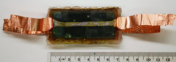
An interdisciplinary team of researchers at Vanderbilt University have developed a way to combine the photosynthetic protein that converts light into electrochemical energy in spinach with silicon, the material used in solar cells, in a fashion that produces substantially more electrical current than has been reported by previous “biohybrid” solar cells.
“This combination produces current levels almost 1,000 times higher than we were able to achieve by depositing the protein on various types of metals. It also produces a modest increase in voltage,” said David Cliffel, associate professor of chemistry, who collaborated on the project with Kane Jennings, professor of chemical and biomolecular engineering.
“If we can continue on our current trajectory of increasing voltage and current levels, we could reach the range of mature solar conversion technologies in three years.”
The researchers’ next step is to build a functioning PS1-silicon solar cell using this new design. Jennings has an Environmental Protection Agency award that will allow a group of undergraduate engineering students to build the prototype. The students won the award at the National Sustainable Design Expo in April based on a solar panel that they had created using a two-year old design. With the new design, Jennings estimates that a two-foot panel could put out at least 100 milliamps at one volt – enough to power a number of different types of small electrical devices.

Harnessing the power of spinach

More than 40 years ago, scientists discovered that one of the proteins involved in photosynthesis, called Photosystem 1 (PS1), continued to function when it was extracted from plants like spinach. Then they determined PS1 converts sunlight into electrical energy with nearly 100 percent efficiency, compared to conversion efficiencies of less than 40 percent achieved by manmade devices. This prompted various research groups around the world to begin trying to use PS1 to create more efficient solar cell

Another potential advantage of these biohybrid cells is that they can be made from cheap and readily available materials, unlike many microelectronic devices that require rare and expensive materials like platinum or indium. Most plants use the same photosynthetic proteins as spinach. In fact, in another research project Jennings is working on a method for extracting PS1 from kudzu.
Since the initial discovery, progress has been slow but steady. Researchers have developed ways to extract PS1 efficiently from leaves. They have demonstrated that it can be made into cells that produce electrical current when exposed to sunlight. However, the amount of power that these biohybrid cells can produce per square inch has been substantially below that of commercial photovoltaic cells.
Another problem has been longevity. The performance of some early test cells deteriorated after only a few weeks. In 2010, however, the Vanderbilt team kept a PS1 cell working for nine months with no deterioration in performance.

“Nature knows how to do this extremely well. In evergreen trees, for example, PS1 lasts for years,” said Cliffel. “We just have to figure out how to do it ourselves.”

Secret is “doping” silicon

The Vanderbilt researchers report that their PS1/silicon combination produces nearly a milliamp (850 microamps) of current per square centimeter at 0.3 volts. That is nearly two and a half times more current than the best level reported previously from a biohybrid cell. The reason this combo works so well is because the electrical properties of the silicon substrate have been tailored to fit those of the PS1 molecule. This is done by
implanting electrically charge atoms in the silicon to alter its electrical properties: a process called “doping.” In this case, the protein worked extremely well with silicon doped with positive charges and worked poorly with negatively doped silicon.
To make the device, the researchers extracted PS1 from spinach into an aqueous solution and poured the mixture on the surface of a p-doped silicon wafer. Then they put the wafer in a vacuum chamber in order to evaporate the water away leaving a film of protein. They found that the optimum thickness was about one micron, about 100 PS1 molecules thick.

Protein alignment

When a PS1 protein exposed to light, it absorbs the energy in the photons and uses it to free electrons and transport them to one side of the protein. That creates regions of positive charge, called holes, which move to the opposite side of the protein.
In a leaf, all the PS1 proteins are aligned. But in the protein layer on the device, individual proteins are oriented randomly. Previous modeling work indicated that this was a major problem. When the proteins are deposited on a metallic substrate, those that are oriented in one direction provide electrons that the metal collects
while those that are oriented in the opposite direction pull electrons out of the metal in order to fill the holes that they produce. As a result, they produce both positive and negative currents that cancel each other out to leave a very small net current flow.
The p-doped silicon eliminates this problem because it allows electrons to flow into PS1 but will not accept them from protein. In this manner, electrons flow through the circuit in a common direction.

source : http://news.vanderbilt.edu/2012/09/spinach-power-a-major-boost/photo-2-241_20120828170545-biohybridcell600/


read more