-->

TEST

Friday 28 December 2012

Strange behavior: new study exposes living cells to synthetic protein

One approach to understanding components in living organisms is to attempt to create them artificially, using principles of chemistry, engineering and genetics. A suite of powerful techniques—collectively referred to as synthetic biology—have been used to produce self-replicating molecules, artificial pathways in living systems and organisms bearing synthetic genomes.
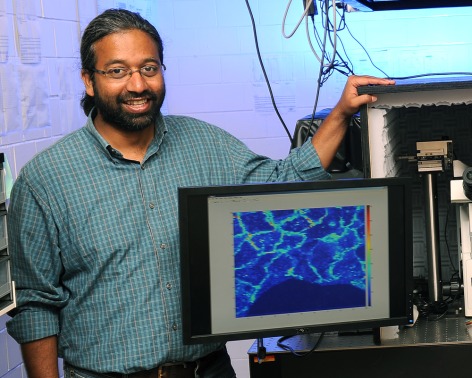
In a new twist, John Chaput, a researcher at Arizona State University’s Biodesign Institute and colleagues at the Department of Pharmacology, Midwestern University, Glendale, AZ have fabricated an artificial protein in the laboratory and examined the surprising ways living cells respond to it.
“If you take a protein that was created in a test tube and put it inside a cell, does it still function,” Chaput asks. “Does the cell recognize it? Does the cell just chew it up and spit it out?”  This unexplored area represents a new domain for synthetic biology and may ultimately lead to the development of novel therapeutic agents.
The research results, reported in the advanced online edition of the journal ACS Chemical Biology, describe a peculiar set of adaptations exhibited by Escherichia coli bacterial cells exposed to a synthetic protein, dubbed DX. Inside the cell, DX proteins bind with molecules of ATP, the energy source required by all biological entities.
“ATP is the energy currency of life,” Chaput says. The phosphodiester bonds of ATP contain the energy necessary to drive reactions in living systems, giving up their stored energy when these bonds are chemically cleaved. The depletion of available intracellular ATP by DX binding disrupts normal metabolic activity in the cells, preventing them from dividing, (though they continue to grow).
After exposure to DX, the normally spherical E. coli bacteria develop into elongated filaments. Within the filamentous bacteria, dense intracellular lipid structures act to partition the cell at regular intervals along its length (see figure 1). These unusual structures, which the authors call endoliposomes, are an unprecedented phenomenon in such cells.
“Somewhere along the line of this filamentation, other processes begin to happen that we haven’t fully understood at the genetic level, but we can see the results phenotypically,” Chaput says. “These dense lipid structures are forming at very regular regions along the filamented cell and it looks like it could be a defense mechanism, allowing the cell to compartmentalize itself.”  This peculiar adaptation has never been observed in bacterial cells and appears unique for a single-celled organism.
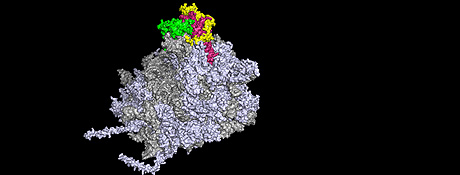
Producing a synthetic protein like DX, which can mimic the elaborate folding characteristics of naturally occurring proteins and bind with a key metabolite like ATP is no easy task.  As Chaput explains, a clever strategy known as mRNA display was used to produce, fine-tune and amplify synthetic proteins capable of binding ATP with high affinity and specificity, much as a naturally occurring ATP-binding protein would.
The depletion of ATP in cells of the bacterium Escherichia coli causes them to
transition to a filamentous state and form dense lipid structures known as endoliposomes.
The structures can be clearly observed in these transmission electron micrographs of increasing magnification.

First, large libraries of random sequence peptides are formed from the four nucleic acids making up DNA, with each strand measuring around 80 nucleotides in length. These sequences are then transcribed into RNA with the help of an enzyme—RNA polymerase.  If a natural ribosome is then introduced, it attaches to the strand and reads the random sequence RNA as though it was a naturally-occurring RNA, generating a synthetic protein as it migrates along the strand. In this way, synthetic proteins based on random RNA sequences can be generated. 
Exposing the batch of synthetic proteins to the target molecule and extracting those that bind can then select for ATP-binding proteins.  But as Chaput explains, there’s a problem: “The big question is how do you recover that genetic information? You can’t reverse transcribe a protein back into DNA. You can’t PCR amplify a protein. So we have to do all these molecular biology tricks.”
The main trick involves an earlier step in the process. A molecular linker is chemically attached to the RNA templates, such that each RNA strand forms a bond with its newly translated protein. The mRNA-protein hybrids are exposed to selection targets (like ATP) over consecutive rounds of increasing stringency. After each round of selection, those library members that remain bound to the target are reverse-transcribed into cDNA (using their conveniently attached RNA messages), and then PCR amplified.
In the current study, E. coli cells exposed to DX transitioned into a filamentous form, which can occur naturally when such cells are subject to conditions of stress. The cells display low metabolic activity and limited cell division, presumably owing to their ATP-starved condition.
The study also examined the ability of E. coli to recover following DX exposure. The cells were found to enter a quiescent state known as viable but non-culturable (VBNC), meaning that they survived ATP sequestration and returned to their non-filamentous state after 48 hours, but lost their reproductive capacity. Further, this condition was difficult to reverse and seems to involve a fundamental reprogramming of the cell.
In an additional response to DX, the filamentous cells form previously undocumented structures, which the authors refer to as endoliposomes. These dense lipid concentrations, spanning the full width of the filamented E. coli, segment the cells into distinct compartments, giving the cells a stringbean-like appearance under the microscope.
The authors speculate that this adaptation may be an effort to maintain homeostasis in regions of the filamentous cell, which have essentially been walled off from the intrusion of ATP-depleting DX. They liken endoliposomes to the series of water-tight compartments found in submarines which are used to isolate damaged sections of the ship and speculate that DX-exposed cells are partitioning their genetic information into regions where it can be safely quarantined. Such self-compartmentalization is known to occur in some eukaryotic cells, but has not been previously observed in prokaryotes like E. coli.
The research indicates that there is still a great deal to learn about bacterial behavior and the repertoire of responses available when such cells encounter novel situations, such as an unfamiliar, synthetic protein. The study also notes that many infectious agents rely on a dormant state, (similar to the VBNC condition observed in the DX-exposed E. coli),  to elude detection by antibiotics. A better understanding of the mechanisms driving this behavior could provide a new approach to targeting such pathogens.
The relative safety of E. coli as a model organism for study may provide a fruitful tool for more in-depth investigation of VBNC states in pathogenic organisms. Further, given ATP’s central importance for living organisms, its suppression may provide another avenue for combating disease. One example would be an engineered bacteriophage capable of delivering DX genes to pathogenic organisms.
 source:http://www.biodesign.asu.edu/news/strange-behavior-new-study-exposes-living-cells-to-synthetic-protein-
read more

Thursday 27 December 2012

New Data Challenge Old Views About Evolution of Early Life

A research team led by biogeochemists at the University of California, Riverside has tested a popular hypothesis in paleo-ocean chemistry, and proved it false.
The fossil record indicates that eukaryotes — single-celled and multicellular organisms with more complex cellular structures compared to prokaryotes, such as bacteria — show limited morphological and functional diversity before 800-600 million years ago. Many researchers attribute the delayed diversification and proliferation of eukaryotes, which culminated in the appearance of complex animals about 600 million years ago, to very low levels of the trace metal zinc in seawater.
As it is for humans, zinc is essential for a wide range of basic cellular processes.  Zinc-binding proteins, primarily located in the cell nucleus, are involved in the regulation of gene transcription.
Organic-rich shale samples, such as these
 from the 2.5-billion-year-old Mount McRae
 Shale from Western Australia, were
analyzed for their zinc contents. The results
 confirm that the early ocean was not
 Zn-lean and that other controls must be
invoked to explain the protracted
 appearance and proliferation of eukaryotic
 life.
Eukaryotes have increasingly incorporated zinc-binding structures during the last third of their evolutionary history and still employ both early- and late-evolving zinc-binding protein structures.  Zinc is, therefore, of particular importance to eukaryotic organisms.  And so it is not a stretch to blame the 1-2-billion-year delay in the diversification of eukaryotes on low bioavailability of this trace metal.
But after analyzing marine black shale samples from North America, Africa, Australia, Asia and Europe, ranging in age from 2.7 billion years to 580 million years old, the researchers found that the shales reflect high seawater zinc availability and that zinc concentrations during the Proterozoic (2.5 billion to 542 million years ago) were similar to modern concentrations. Zinc, the researchers posit, was never biolimiting.
Study results appear online Dec. 23 in Nature Geoscience.
“We argue that the concentration of zinc in ancient marine black shales is directly related to the concentrations of zinc in seawater and show that zinc is abundant in these rocks throughout Earth’s history,” said Clint Scott, the first author of the research paper and a former UC Riverside graduate student. “We found no evidence for zinc biolimitation in seawater.”
Scott, now a research geologist with the U.S. Geological Survey, explained that the connection between zinc limitation and the evolution of eukaryotes was based largely on the hypothesis that Proterozoic oceans were broadly sulfidic. Under broadly sulfidic conditions, zinc should have been scarce because it would have rapidly precipitated in the oceans, he explained.
“However, a 2011 research paper in Nature also published by our group at UCR demonstrated that Proterozoic oceans were more likely broadly ferruginous — that is, low in oxygen and iron-rich — and that sulfidic conditions were more restricted than previously thought,” said Scott, who performed the research in the lab of Timothy Lyons, a professor of biogeochemistry and the principal investigator of the research project.
The research team argues that ferruginous deep oceans, combined with large hydrothermal fluxes of zinc via volcanic activity on the seafloor, maintained high levels of dissolved zinc throughout the oceans and provided a relatively stable marine reservoir of the trace metal over the past 2.7 billion years.
“The key challenge in understanding the early evolution of life is recognizing the environmental conditions under which that life first appeared and diversified,” Lyons said. “We have taken a very direct approach that specifically tracks the availability of essential micronutrients, and, to our surprise, zinc supplies in ancient seawater were much higher and less variable than previously imagined.
“We can imagine for the first time,” he quipped, “that zinc supplements were not on the shopping lists of our early eukaryotic ancestors, and so we better find another reason to explain the mysterious delay in their rise in the ocean.”
SOURCE:http://ucrtoday.ucr.edu/10927
read more

Thursday 20 December 2012

GEIC deadline extended to 31st December


GEIC

    GBioFin Entrepreneurship and Innovation Certificate
    NOW IT’S YOUR TURN TO BE THE NEXT KIRAN MAZUMDAR!!!!!!!!!!!


We come across many students who have lots of business ideas during their graduation days, about opening their own business and a dream of being the Boss /CEO of their own company. But with time, these ideas get cornered and a person ends up finding a good job or takes up higher studies etc, but very few have the courage to start their own venture.

This happens due to many reasons like
a) Lack of knowledge like about how to go into the business
b) Improper direction as to what are the requirements for starting a business 
c) No guidance or less courage to start their own venture.

GBioFin (Gate to Biotech Industries, Organizations and Foundations in India) proudly announces the launching of GEIC (GBioFin Entrepreneurship and Innovation Certificate), a program specialized for entrepreneurship learning.
GEIC is 3 months Online Program started for promoting Biotechnology and Life Sciences in Entrepreneurship field. GEIC aims to provide all the Information regarding Entrepreneurship, Innovation, Research, and Intellectual Property Rights and so on.

It’s a 3-month online course, in which we will give you a larger and a practical picture of the steps / requirements that you need to go through before starting your own venture.
This 3 month program  will give you an edge over other students. Usually, students do  this kind of course after graduating from college so getting this know-how at the college level will at least save 1.5 years of your life.

Programme Includes-
     1. Online Interactive Sessions
     2. Online best course material and module notes (based on patterns from Cambridge and Oxford)
     3. E-Copies of Quarterly Magazine(Biotech Rings) for all the registered  students.
     4. Launch of a student Educational Magazine (E-Copy) for GEIC enrolled Students(including Articles given as assignment for GEIC)
     5.  Interaction with the advisor assigned for the GEIC through email
     6. GEIC Certificate will be sent at the home addresses of each candidate.
     7. Entry in the GBioFin Events with Heavy Discount/ Free Entry in some cases
     8. Gate100 Online Study materials for all.

And many more…

Registration Fees= 1000 /-

To register for GEIC please visit our website www.biofin.net, all the information and details are provided on the homepage.

Contact us:-
Shah Vaibhav-   09769253423
Co-Founder GBioFin Biotechnology Services

read more

TATA First Dot Student Start-up Award


APPEAL

We have been nominated for Tata first dot Student Start-up Award    Powered by NEN
VOTE for us  ( GBioFin Biotechnology Services) at Tata first dot to BRING THE CHANGE

Get yourself registered  and VOTE for Biotech.
Remember "VOTE" is different from the "LIKE" option. So do cast your vote. AND BRING THE CHANGE !!!!!!!
3 Steps to Cast your VOTE :-
1)Click on the link http://tatafirstdot.com/nomination/gbiofin
2)Click on the Thumbs up Option
3)Sign Up and Like again on the FB Link ( confirmation link will be send to your email id)
4) After the Click on the confirmation link your will be able to CAST YOUR VOTE
5) After you VOTE , make sure your Vote is registered.
- Make sure you VOTE and not just Like 

We have got 4,195 members on Facebook group. We can win this. Please vote.

Come Join Us in our Movement to BRING THE CHANGE !!!!!!!

read more

CSIR UGC NET(Life Sciences)


This post is dedicated to all the NET aspirants who will be taking the test this Sunday i.e. the 23rd December 2012. 
We hope you are all geared up and ready to rock the test.

CSIR NET exam is really a tough one and thousands of aspirants desire to crack it. It needs a thorough preparation which you all must be done with by now and a well planned strategy for taking the exam.
Here in this post we will be providing you some tips about CSIR UGC NET.


1. The first suggestion to all is to keep a check on the CSIR HRDG website always, even after you have taken the exam.
For instance the timings of the exams was changed as given here.

2. Try doing a lot of question papers of the previous NET exams, as many as possible. This helps in improving your speed which is a crucial factor when one is attempting the questions in very limited time.
In addition to that, it helps in preparing a strategy which you are most likely to follow in your exam to manage your time between questions (not all questions need to be attempted) and obtain the maximum efficiency and as a consequence the maximum marks.

3. Work on your strong topics very exhaustively. Since only 15/20, 35/50 and 25/75 questions need to be done, it will help you to decide the questions quickly thus saving a lot of time. In other words you should have complete knowledge of the topic you are very good at.

4. Never answer the questions randomly. Answer only if you are sure of the answer.
Sometimes categorizing the questions into 3 categories, absolutely sure ones, doubtful ones and the ones with no idea; is helpful. This way you can attempt the first category and only if the required number of questions have not been attempted then only you should try the next category questions.

Last of all, GBioFin wishes "All the Best" to all the candidates.


Just for reference sake:
Syllabus change 
Some Books for reference :

1. Upkars CSIR-UGC/NET/JRF/SLET Life Sciences by Dr.A.P.Singh Kumar Pushkara
2. Truemans UGC-CSIR JRF/NET Life Sciences by Pramod Singh

read more

Wednesday 19 December 2012

Designing Influenza Immunogen


Influenza HA is the primary target of neutralizing antibodies during infection, and its sequence undergoes genetic drift and shift in response to immune pressure. The receptor binding HA1 subunit of HA shows much higher sequence variability relative to the metastable, fusion-active HA2 subunit, presumably because neutralizing antibodies are primarily targeted against the former in natural infection. We have designed an HA2-based immunogen using a protein minimization approach that incorporates designed mutations to destabilize the low pH conformation of HA2. The resulting construct (HA6) was expressed in Escherichia coli and refolded from inclusion bodies. Biophysical studies and mutational analysis of the protein indicate that it is folded into the desired neutral pH conformation competent to bind the broadly neutralizing HA2 directed monoclonal 12D1, not the low pH conformation observed in previous studies. HA6 was highly immunogenic in mice and the mice were protected against lethal challenge by the homologous A/HK/68 mouse-adapted virus. An HA6-like construct from another H3 strain (A/Phil/2/82) also protected mice against A/HK/68 challenge. Regions included in HA6 are highly conserved within a subtype and are fairly well conserved within a clade. Targeting the highly conserved HA2 subunit with a bacterially produced immunogen is a vaccine strategy that may aid in pandemic preparedness.


Read the full study here.
A summary is given here.


read more

Sunday 2 December 2012

Hiyoshi Internship Program 2013

Hiyoshi Corporation, Omihachiman, Japan and ABK-AOTS DOSOKAI, Tamilnadu Centre are jointly organizing two months stipendiary International Internship Program’2013 at Japan for three students from the Engineering Colleges / Universities of India.

The students can be from Biotechnology, Environmental Science & Chemical Engineering and they should have consistently scored 75% of marks throughout.

Preferences will be given to those students who are having knowledge in basic Japanese Language skill.


Students who are perusing Third Year of Engineering or 1st yr of post graduation can apply for this program

Terms & Conditions and Application Form for the Training at Hiyoshi Corporation, Japan is in below link.

Internship details

Application form


read more

Monday 26 November 2012

Capturing living cells in micro pyramids

Cells moving into the pyramids
A field full of pyramids, but on a micro scale. Each of the pyramids hides a living cell. Thanks to 3D micro- and nano scale fabrication, promising new applications can be found. One of them is applying the micro pyramids for cell research: thanks to the open ‘walls’ of the pyramids, the cells interact. Scientists of the research institutes MESA+ and MIRA of the University of Twente in The Netherlands present this new technology and first applications in Small journal of the beginning of December.
Most of the cell studies take place in 2D: this is not a natural situation, because cells organize themselves in another way than in the human body. If you give the cells room to move in three dimensions, the natural situation is approached in a better way while capturing them in an array. This is possible in the ‘open pyramids’ fabricated in the NanoLab of the MESA+ Institute for Nanotechnology of the University of Twente.
Tiny corner remains filled
The cleanroom technology applied for this, has been discovered by coincidence and is now called ‘corner lithography’. If you join a number of flat silicon surface in a sharp corner, it is possible to deposit another material on them. After having removed the material, however, a small amount of material remains in the corner. This tiny tip can be used for an Atomic Force Microscope, or, in this case, for forming a micro pyramid.
Catching cells
Chondrocyte captured inside a micro pyramid, interacting with its neighbours
In cooperation with UT’s MIRA Institute for Biomedical Technology and Technical Medicine, the nanoscientists have explored the possibilities of applying the pyramids as ‘cages’ for cells. First experiments with polystyrene balls worked out well. The next experiments involved capturing chondrocytes, cells forming cartilage. Moved by capillary fluid flow, these cells automatically ‘fall’ into the pyramid through a hole at the bottom. Soon after they settle in their 3D cage, cells begin to interact with cells in adjacent pyramids. Changes in the phenotype of the cell can now be studied in a better way than in the usual 2D situation. It is therefore a promising tool to be used in for example tissue regeneration research.
read more

Thursday 15 November 2012

Solving the mystery of ageing

Why do we get older? When do we die and why? Is there a life without ageing? For centuries, science has been fascinated by these questions. Now researchers from Kiel (Germany) have examined why the polyp Hydra is immortal – and unexpectedly discovered a link to ageing in humans. The study carried out by Kiel University together with the University Medical Center Schleswig-Holstein (UKSH) will be published this week in the Proceedings of the National Academy of Sciences of the United States of America (PNAS). It was funded by the German Research Foundation DFG.

Hydra – mysteriously immortal
The tiny freshwater polyp Hydra does not show any signs of ageing and is potentially immortal. There is a rather simple biological explanation for this: these animals exclusively reproduce by budding rather than by mating. A prerequisite for such vegetative-only reproduction is that each polyp contains stem cells capable of continuous proliferation. Without these stem cells, the animals could not reproduce any more. Due to its immortality, Hydra has been the subject of many studies regarding ageing processes for several years.

Ageing in humans
Caption:A gene sequence is injected into an embryo of Hydra.
Copyright/Foto: CAU/Wittlieb
Human longevity gene discovered in Hydra
„Surprisingly, our search for the gene that causes Hydra to be immortal led us to the so-called FoxO gene“, says Anna-Marei Böhm, PhD student and first author of the study. The FoxO gene exists in all animals and humans and has been known for years. However, until now it was not known why human stem cells become fewer and inactive with increasing age, which biochemical mechanisms are involved and if FoxO played a role in ageing. In order to find the gene, the research group isolated Hydra’s stem cells and then screened all of their genes.

Immortality mechanism of Hydra revealed
The Kiel research team examined FoxO in several genetically modified polyps: Hydra with normal FoxO, with inactive FoxO and with enhanced FoxO. The scientists were able to show that animals without FoxO possess significantly fewer stem cells. Interestingly, the immune system in animals with inactive FoxO also changes drastically. „Drastic changes of the immune system similar to those observed in Hydra are also known from elderly humans“, explains Philip Rosenstiel of the Institute of Clinical Molecular Biology at UKSH, whose research group contributed to the study.

FoxO makes human life longer, too
„Our research group demonstrated for the first time that there is a direct link between the FoxO gene and ageing“, says Thomas Bosch from the Zoological Institute of Kiel University, who led the Hydra study. Bosch continues: „ FoxO has been found to be particularly active in centenarians – people older than one hundred years – which is why we believe that FoxO plays a key role in ageing – not only in Hydra but also in humans“. However, the hypothesis cannot be verified on humans, as this would require a genetic manipulation of humans. Bosch stresses however that the current results are still a big step forward in explaining how humans age. Therefore the next step must be to study how the longevity gene FoxO works in Hydra, and how environmental factors influence FoxO activity.

Without stem cells we all die
Scientifically, the study has two major conclusions: On the one hand it confirms that the FoxO gene plays a decisive role in the maintenance of stem cells. It thus determines the life span of animals – from cnidarians to humans. On the other hand, the study shows that ageing and longevity of organisms really depend on two factors: the maintenance of stem cells and the maintenance of a functioning immune system.
source:http://www.uni-kiel.de/aktuell/pm/2012/2012-332-foxogen-e.shtml
read more

How Do Cells Tell Time? Scientists Develop Single-Cell Imaging to Watch the Cell Clock

A new way to visualize single-cell activity in living zebrafish embryos has allowed scientists to clarify how cells line up in the right place at the right time to receive signals about the next phase of their life.
Under normal circumstances in zebrafish embryos, cells oscillate in synchrony
with their neighbors as they prepare to make segments that later become
muscle and vertebrae. When a color map (top left corner) is used to indicate the
 phase of oscillation in each cell at any fixed snapshot of time, with cool colors
representing the peak of the gene activation wave and warm colors the lower
levels of activation, it is evident in the top image that neighboring cells are in a
similar phase, or transitioning smoothly to the next phase. However, in embryos
 lacking a powerful messaging system called Notch signaling, that synchrony is
lost. In the bottom map, cells in mutant embryos that lack the Notch signal are
oscillating, but the random assortment of colors without smooth transitions
 shows that Notch is required to synchronize the oscillations in neighboring cells.
Images courtesy of Ohio State University
Scientists developed the imaging tool in single living cells by fusing a protein defining the cells’ cyclical behavior to a yellow fluorescent protein that allows for visualization. Zebrafish embryos are already transparent, but with this closer microscopic look at the earliest stages of life, the researchers have answered two long-standing questions about how cells cooperate to form embryonic segments that later become muscle and vertebrae.
Though these scientists are looking at the molecular “clock” that defines the timing of embryonic segmentation, the findings increase understanding of cyclical behaviors in all types of cells at many developmental stages – including problem cells that cause cancer and other diseases. Understanding how to manipulate these clocks or the signals that control them could lead to new ways to treat certain human conditions, researchers say.
Embryonic cells go through oscillating cycles of high and low signal reception in the process of making segmented tissue, and gene activation by the groups of cells must remain synchronized for the segments to form properly. One of a handful of powerful messaging systems in all vertebrates is called the Notch signaling pathway, and its precise role in this oscillation and synchronization has been a mystery until now.
In this study, the researchers confirmed that the cells must receive the Notch signal to maintain synchronization with nearby cells and form segments that will become tissue, but the cells can activate their genes in oscillating patterns with or without the signal.
“For the first time, this nails it,” said Sharon Amacher, professor of molecular genetics at Ohio State University and lead author of the study. “This provides the data that cells with disabled Notch signaling can oscillate just fine, but what they can’t do is synchronize with their neighbors.”
The imaging also allowed Amacher and colleagues to determine that cell division, called mitosis, is not a random event as was once believed. Instead, division tends to occur when neighboring cells are at a low point of gene activation for signal reception – suggesting mitosis is not as “noisy,” or potentially disruptive, as it was previously assumed.
 The study is published in the November issue of the journal Developmental Cell.
Amacher’s work focuses on the creation of these tissue segments, called somites, in the mesoderm of zebrafish embryos – this region gives rise to the ribs, vertebrae and muscle in all vertebrates, including humans.
“This early process of segmentation is really important for patterning a lot of subsequent developmental events – the patterning of the nervous system and the vasculature, much of that depends on this clock ensuring that early development happens properly,” Amacher said.
Unlike the well-known 24-hour Circadian clock, however, the activities of cells at the earliest stages of development can occur within a matter of minutes – which makes their clocks very challenging to study.

Experts in tissue segmentation liken the oscillating cycle of gene activation and de-activation that cells go through before they form somites to the wave that fans perform in a stadium. According to the segmentation clock, genes are turned on, proteins are made, proteins then inhibit gene activation, and so on, and the pattern repeats until all necessary somites are formed. Neighbor cells must be in sync with each other just as sports fans in the same section must stand and sit at the same time to effectively form a wave.
Zebrafish somites form every 30 minutes, meaning that during any one cycle of the wave, a cell is engaged in making protein for only about five minutes. To generate the imaging tool, researchers linked a yellow fluorescent protein to a cyclic protein known to have a short lifespan. The resulting short-lived fluorescent fusion protein allowed Amacher and colleagues to look at single cells along with their neighbors to observe how they stayed synchronized as they did the wave.
Researchers in this field had previously thought that the Notch signaling pathway may be needed to start the clock in these cyclic genes, though conflicting data had shown that the clock could run without the signal.
Amacher’s imaging showed that, indeed, Notch was required only to maintain synchronization, but not to start the oscillating clock. She and colleagues tested this idea by combining the imaging tool with three mutant cell types with disabled Notch signals. Cells in all three mutants could oscillate, but not in a synchronized fashion, explaining how they failed to form segments in the way that cells receiving the Notch signal could.
Defects in Notch signaling are associated with human congenital developmental disorders characterized by malformed ribs and vertebrae, suggesting this work offers insight into potential therapies to prevent these defects.
The researchers next sought to determine whether cell division interrupted the synchrony needed for creation of the segments. Mitosis, occurring among 10 to 15 percent of embryonic cells at any one time, is considered a source of biological “noise” because when cells divide, they stop activating genes. If division were happening randomly, as previously thought, instead of in a pattern, the very cell division needed for organism growth could also disrupt clock synchrony, creating problems that segmenting organisms would have to overcome.
The study showed, however, that most cells divided when their neighbors were at a low point of gene activation – at the bottom of a wave – suggesting that cell division doesn’t occur at random. The study team noted that the two daughter cells created from a fresh division are more tightly synchronized with each other than are any other cell neighbors in the area.
Under normal conditions, these two daughters resynchronize with their neighbors in short order. In embryos lacking Notch signaling, newly divided daughters appeared as a pair of tightly synchronous cells in a largely asynchronous sea, showing that oscillation could resume without the signaling pathway. Without Notch, the daughter cells gradually drifted out of synchrony, becoming like their asynchronous neighbors.
Amacher said these findings could be incorporated into models of developmental cell behavior to further advance cell biology research.
“Most of our tissues and organs are not made up of the same types of cells. They have different jobs. So you don’t want them to respond identically to every signal; you want them to have different responses,” she said. “We need to understand systems like this that help cells not only interpret the signals in their environment, but do the right thing when they get that signal.”
source:http://researchnews.osu.edu/archive/cellclock.htm
read more

GBioFin Entrepreneurship and Innovation Certificate

GBioFin proudly announces the launching of GEIC (GBioFin Entrepreneurship and Innovation Certificate).

GEIC is 3 months Online Programme started for Promoting Biotechnology and Life-Sciences with its scope in Entrepreneurship.GEIC aims to provide all the Information regarding Entrepreneurship, Innovation, Research, Intellectual Property Rights and so on.

Program Includes-
1-Online Interactive Sessions
2-Online best course material and module notes (having comparison with Cambridge and Oxford) with assignments
3- Launch of a student Educational Magazine E-Copy for GEIC enrolled Students (including Articles given as assignment for GEIC)
4-Interaction with the advisors assigned for the GEIC through email
5-GEIC Certificate will be sent at the home addresses of each candidate through post
6-Student Membership free for 1 year worth Rs.350 which includes the following:-
  a) Free Access & heavy discounts in Honorable Programs like GECP, GATE100, where they will         get and entry free of cost
  b) Latest jobs updates and first preferences to them for the jobs referred by GBioFin
  c) Heavy discounts in the Conferences conducted in Association GBioFin
  d) Yearly Subscription of e-Magazine free worth Rs.250
And much more ……….

To register for GEIC please visit our website www.biofin.net or register at geic@biofin.net, the information will be provided there very soon …
read more

Tuesday 13 November 2012

Jellyfish-inspired device that rapidly and efficiently captures cancer cells from blood samples could enable better patient monitoring

Cells traveling through a microfluidic device can be trapped
by strands of DNA (green).
Image: Suman Bose and Chong Shen
Tumor cells circulating in a patient’s bloodstream can yield a great deal of information on how a tumor is responding to treatment and what drugs might be more effective against it. But first, these rare cells have to be captured and isolated from the many other cells found in a blood sample.

Many scientists are now working on microfluidic devices that can isolate circulating tumor cells (CTCs), but most of these have two major limitations: It takes too long to process a sufficient amount of blood, and there is no good way to extract cancer cells for analysis after their capture.

A new device from researchers at MIT and Brigham and Women’s Hospital overcomes those obstacles. Inspired by the tentacles of a jellyfish, the team coated a microfluidic channel with long strands of DNA that grab specific proteins found on the surfaces of leukemia cells as they flow by. Using this strategy, the researchers achieved flow rates 10 times higher than existing devices — fast enough to make the systems practical for clinical use.

Using this technology, described in this week’s issue of the Proceedings of the National Academy of Sciences, doctors could monitor cancer patients to determine whether their treatment is working.

“If you had a rapid test that could tell you whether there are more or less of these cells over time, that would help to monitor the progression of therapy and progression of the disease,” says Jeff Karp, an associate professor of medicine at Harvard Medical School and co-director of the Center for Regenerative Therapeutics at Brigham and Women’s Hospital in Boston.

This type of device could also enable personalized treatments: Once cells are isolated from a patient, doctors could test different drugs on them to determine which are most effective.

DNA ‘tentacles’

The number of CTCs found in a milliliter of a particular patient’s blood can range from just a few to several thousand. To isolate those rare cells, researchers have tried building microfluidic channels dotted with antibodies specific to a protein found on the target cells. However, because the antibodies only extend tens of nanometers from the bottom of the channel, the capture of cells by the antibodies is slow.

To extend the reach of the capture molecules, Karp and Karnik’s team mimicked the tentacles of jellyfish, creating long strands of repeating DNA sequences. Those sequences, known as aptamers, target a protein found in large numbers on leukemia cells.

The DNA strands are attached to a microchannel with a herringbone pattern on its floor. Those patterned ridges cause the blood to swirl as it flows through the channel, improving the chances that individual cells will come into contact with the tentacles, which extend hundreds of microns into the channel. This allows the researchers to increase the rate of blood flow.

“Normally what happens at high flow rates is the cells don’t really come close to the surface, and it’s very challenging to capture the target cells,” Karnik says. “But this combination of these herringbone grooves to mix the solution and bring the cells into contact with surfaces, plus having aptamers that are sticking out into the solution, enables very high capture rates at very high flow rates.”

Flow rates in the new device are 10 times higher than those reported for previous devices, and the system can capture 60 to 80 percent of the target cells. In the current model, which measures 1 square centimeter, the flow rate is 1 milliliter per hour. By making the device larger, the researchers say they could boost the flow rate to 100 milliliters of blood per hour — fast enough to rapidly process the 10- to 20-milliliter samples that would be needed to get an accurate CTC count from an individual patient.

Because the “tentacles” are made of DNA, they can easily be cleaved with enzymes, freeing the captured cells for further analysis.

Peter Kuhn, an associate professor of cell biology at the Scripps Research Institute, says this technology is an interesting alternative to existing approaches to capture rare cells, adding that further testing is needed to validate the technology’s usefulness.

“This field needs many different avenues for rare-cell identification to be explored,” Kuhn says. “These technologies can provide the key enablers for studying disease in humans.”

Minimally invasive

Devices that capture CTCs could offer a better alternative to sampling bone marrow in determining whether cancer treatment is working in a leukemia patient.

“If one could improve the sensitivity of detection in blood, then this approach may enable a transition from isolating marrow to isolating blood, which is much less invasive and you can do it more often. It could change the paradigm for how residual disease is detected,” Karp says.

“The beauty of this technology is its versatility,” Zhao says. “You can easily modify the length and density of the DNA chains; you can include different sequences in the DNA to capture different types of cells.”

This feature makes it a platform technology that can be broadly applied in the clinic and research laboratories. For instance, another possible application is capturing fetal cells, which are very rare in a pregnant woman’s bloodstream. Analyzing these cells could help doctors perform prenatal diagnostic tests for a range of diseases using an approach that is far less invasive than amniocentesis.

The researchers are now working on adapting the DNA strands to target other molecules, such as receptors found on the surfaces of cells dislodged from solid tumors
source:http://web.mit.edu/newsoffice/2012/capturing-rare-cancer-cells-1112.html
read more

Sunday 11 November 2012

First ‘snapshots’ of the electronic structure of a manganese complex related to water-splitting in photosynthesis

Together with a large international research team, Johannes Messinger of Umeå University in Sweden has taken another step toward an understanding of photosynthesis and developing artificial photosynthesis. With a combination of a x-ray free-electron laser and spectroscopy, the team has managed to see the electronic structure of a manganese complex, a chemical compound related to how photosynthesis splits water.
 illustration of ultra-short x-ray pulse striking molecules
containing manganese. Illustration: Greg Stewart,
National Accelerator Laboratory at Stanford University
The experiments used the Linac Coherent Light Source (LCLS), which is a free-electron x-ray laser facility at Stanford University in the US. The wavelength of the laser is roughly the same as the breadth of an atom, and each pulse of light lasts 50 femtoseconds (10-15). This is an extremely short interval of time: there are more femtoseconds in one second than there are seconds in a person’s life. Such extremely short wavelengths and short light pulses constitute ideal conditions for imaging chemical reactions with atomic resolution at room temperature while the chemical reactions are ongoing.
The research group has previously used LCLS to perform structural analyses of isolated photosynthesis complexes from plants’ photosystem II at room temperature. Now the group has combined the method with spectroscopy and is the first team to succeed in seeing at LCLS the electronic structure of a manganese complex similar to that found in photosystem II. Manganese is a transitional metal that, together with calcium and oxygen, forms the water-splitting catalyst in photosystem II.
A very simple example of a spectrometer is a prism, which separates sunlight into all the colors of the rainbow. The spectrometer used in this study functions in a similar manner, but with a group of 16 specialized crystals that diffract the x-rays emitted from the sample in resonse of being excited by an x-ray pulse onto a detector array.
To the delight of the scientists, the manganese compounds remained intact long enough for them to observe detailed information about the electronic structure before the compounds were destroyed by the very intense X-ray laser beam.
“Having both structural information and spectroscopic information means that we can much better understand how the structural changes of the whole complex and the chemical changes on the active surface of the catalysts work together to enable the enzymes to perform complex chemical reactions at room temperature,” says Johannes Messinger, professor at the Department of Chemistry at UmeÃ¥ University.
The chemical reaction the research group aims to understand is the splitting of water in photosystem II, as this understanding is also key for developing artificial photosynthesis– that is, for building devices for producing hydrogen from sunlight and water. To be able to exploit sunlight for producing fuels that can be stored and the used when needed would help solve the world’s ever-more acute energy problems.
source:http://www.teknat.umu.se/english/about-the-faculty/news/newsdetailpage/first-snapshots-of-the-electronic-structure-of-a-manganese-complex-related-to-water-splitting-in-photosynthesis.cid202064

read more

Wyss Institute Models a Human Disease in an Organ-on-a-Chip

Researchers at the Wyss Institute for Biologically Inspired Engineering at Harvard University have mimicked pulmonary edema in a microchip lined by living human cells, as reported today in the journal Science Translation Medicine. They used this "lung-on-a-chip" to study drug toxicity and identify potential new therapies to prevent this life-threatening condition.

 The study offers further proof-of-concept that human "organs-on-chips" hold tremendous potential to replace traditional approaches to drug discovery and development.
"Major pharmaceutical companies spend a lot of time and a huge amount of money on cell cultures and animal testing to develop new drugs," says Donald Ingber, M.D., Ph.D., founding director of the Wyss Institute and senior author of the study, "but these methods often fail to predict the effects of these agents when they reach humans."
The lung-on-a-chip device [see video], which the team first described only two years ago, is a crystal clear, flexible polymer about the size of a memory stick that contains hollow channels fabricated using computer microchip manufacturing techniques. Two of the channels are separated by a thin, flexible, porous membrane that on one side is lined with human lung cells from the air sac and exposed to air; human capillary blood cells are placed on the other side with medium flowing over their surface. A vacuum applied to side channels deforms this tissue-tissue interface to re-create the way human lung tissues physically expand and retract when breathing.

Wyss Technology Development Fellow Dongeun Huh, Ph.D., who also holds appointments at Boston Children's Hospital and Harvard Medical School, studied a cancer chemotherapy drug called interleukin-2 -- or IL-2 for short -- in the lung-on-a-chip. A major toxic side effect of IL-2 is pulmonary edema, which is a deadly condition in which the lungs fill with fluid and blood clots.


 When IL-2 was injected into the blood channel of the lung-on-a-chip, fluid leaked across the membrane and two tissue layers, reducing the volume of air in the other channel and compromising oxygen transport -- just as it does in lungs of human patients when it is administered at the equivalent doses and over the same time course. Blood plasma proteins also crossed into the air channel, leading to the formation of blood clots in the air space, as they do in humans treated with IL-2.
But one result came as a surprise.
It turns out the physical act of breathing greatly enhances the effects of IL-2 in pulmonary edema -- "something that clinicians and scientists never suspected before," Ingber says. When the team turned on the vacuum attached to the chip to simulate breathing, it increased fluid leakage more than three-fold when treated with the clinically relevant IL-2 dose, and the Wyss team confirmed that the same response occurs in an animal model of pulmonary edema. This result could suggest that doctors treating patients on a respirator with IL-2 should reduce the tidal volume of air being pushed into the lungs, for example, in order to minimize the negative side effects of this drug.
Combining microfabrication techniques with modern
tissue engineering, lung-on-a-chip offers an in
vitro approach to drug screening by mimicking
the complicated mechanical and biochemical
behaviors of a human lung.
Most exciting for the future of drug testing was the Wyss team's finding that "this on-chip model of human pulmonary edema can be used to identify new potential therapeutic agents in vitro," Ingber says. The pulmonary edema symptoms in the lung-on-a-chip disease model could be prevented by treating the tissues with a new class of drug, a transient receptor potential vanilloid 4 (TRPV4) channel blocker, under development by GlaxoSmithKline (GSK). In a separate study published by the GSK team in the same issue of Science Translation Medicine, the beneficial effects of TRPV4 inhibition in reducing pulmonary edema were independently validated using animal models of pulmonary edema caused by heart failure.
"In just a little more than two years, we've gone from unveiling the initial design of the lung-on-a-chip to demonstrating its potential to model a complex human disease, which we believe provides a glimpse of what drug discovery and development might look like in the future," Ingber says.

source:http://wyss.harvard.edu/viewpressrelease/99/


read more

Monday 5 November 2012

Biochemists Discover New Mechanism in Ribosome Formation

A new mechanism in the formation of ribosomes has been discovered by researchers from the Heidelberg University Biochemistry Center. In an interdisciplinary approach, the Heidelberg scientists, along with colleagues from Switzerland and Japan, describe a heretofore uncharacterised protein that plays a specific role in ribosome assembly in eukaryotes, organisms whose cells contain a cell nucleus. This protein makes sure that specific factors required for ribosome synthesis are transported together, like hitchhikers, into the nucleus to the site of assembly. The results of this research were published in “Science”.
The figure shows the large subunit of the ribosome in its high-resolution 3D
structure. The ribosomal RNA is depicted in grey, the myriad of ribosomal proteins
in blue-grey. The r-protein Rpl5 is shown in yellow, the r-protein Rpl11 in green. The
ruby-coloured area reveals the ribosomal RNA which binds Rpl5 and Rpl11.

Ribosomes, the protein factories of the cell, are macromolecular complexes of ribonucleic acids (RNA) and ribosomal proteins (r-proteins) that are organised in a highly complicated three-dimensional nanostructure. Correct synthesis of ribosomes is critical for the division of all cells and is a process that follows strict rules. In eukaryotes, new ribosomes are formed predominantly in the cell nucleus. Therefore, the r-proteins needed for ribosome formation must travel from the cytoplasm of the cell to a site in the nucleus where the ribosomes are assembled. Until recently it was not clear whether r-proteins that have a similar function and form functional clusters on the ribosome structure are also co-transported into the nucleus.
The researchers have now found a protein that coordinates the co-transport of certain r-proteins in functional clusters into the cell nucleus. This factor is called Symportin1, for synchronised import. “Symportin1 synchronises the import of both the Rpl5 and Rpl11 r-proteins into the cell nucleus and supports their integration into the growing ribosome structure”, explains Prof. Dr. Irmgard Sinning of the Heidelberg University Biochemistry Center (BZH). “It employs a familiar logistical concept from every day life, like picking up a hitchhiker or sharing a taxi with someone headed for the same destination”, says Dr. Gert Bange of the BZH, lead author of the study together with Dr. Dieter Kressler (now of Fribourg University).
source: http://www.uni-heidelberg.de/presse/news2012/pm201211102_ribosomen_en.html
read more

NYU researchers use simulations on TACC, XSEDE supercomputers to understand how some carcinogens evade removal by stabilizing the very DNA they attack

A person doesn't have to go far to find a polycyclic aromatic hydrocarbon (PAH). These carcinogen precursors are inhaled through automobiles exhaust during the morning commute, are present in a drag of cigarette smoke, and are part of any barbequed meal.

Once ingested or inhaled, these big, bulky multi-ringed molecules are converted into reactive carcinogenic compounds that can bind to DNA, sometimes literally bending the double helix out of its normal shape, to form areas of damage called lesions. The damaged DNA can create errors in the genetic code during replication, which may cause cancer-initiating mutations.
It is the job of the nuclear excision repair (NER) system to repair damage caused by PAH lesions by removing the segment of DNA where the lesion is bound and patching up the resulting gap. But some lesions are especially resistant to this repair machinery, making them much more likely to cause mutations than lesions that are promptly repaired.
Models showing the steric hinderance between different lesions (colored yellow and turquoise)and DNA. Steric hinderance causes double helix destabilization and refers to the degree of crowding between different atoms.
A research team at New York University (NYU) has gained new insight on the ability of certain PAH-derived lesions to evade the DNA repair machinery. They found that some lesions stabilize the DNA they damage, making it difficult for a certain repair protein to mark the lesion for repair. Their research was published earlier this year in the February 2012 issue of Biochemistry. More recent articles about NER of DNA lesions from the same group appeared in Nucleic Acids Research in July and August.
"Some lesions cause DNA to be locally destabilized, but there are lesions that actually stabilize the DNA so that the two strands come apart with great difficulty," said Suse Broyde, a biology professor at NYU. "Sometimes they're even more stable than undamaged DNA."
The stability of the DNA double helix is a key feature that determines whether DNA is flagged for repair in the first place by a protein called XPC. The protein patrols the genome looking for weakened areas. When it finds one, it slips a structure called a beta-hairpin between the strands, marking the DNA for NER. But if a lesion makes DNA more stable, the strands become more difficult to separate and the beta hairpin can't signal for repair.
How can molecules as large and disruptive as a PAH stabilize DNA in the first place?


"If you remember from chemistry, every kind of molecule interacts with other molecules through so-called Van der Waals interactions," said Nicholas Geacintov, a chemistry professor at NYU. "The DNA and the carcinogen bound to it also have the same kind of interactions," he said, specifically referring to the PAH-derived lesions that intercalate, or wedge themselves, between DNA base pairs.
The role of Van der Waals forces, which Broyde calls "stacking interactions" for these systems, was made clear through a series of computer simulations performed, analyzed, interpreted and visualized by Yuqin Cai, a post-doctoral senior research scientist in Broyde's lab. "The computer simulations revealed the structural, energetic and dynamic properties of the DNA containing the PAH-derived lesions, " said Broyde, who specializes in providing a mechanistic understanding of complex biological processes using molecular dynamics simulations and other front-line computational approaches.
"You can make movies of the dynamic trajectory which allow you to see the real mobility of the entire system," Broyde said. "You can watch the DNA flexing and the backbone moving dynamically and the carcinogen moving in and out. It's not rigid — you can see its aliveness."

Nuclear Excision Repair of a PAH-derived lesion.
Animation courtesy of Yuqin Cai.

Broyde and her team's simulations revealed that of the six different lesions examined (three chemicals with two different geometric configurations each), those caused by dibenzo[a,l]pyrene, the most tumorigenic PAH investigated, were the most resistant to repair. The five-ringed structure of the carcinogen provided ample stacking opportunities, which stabilized the DNA much better than the four and three-ringed structures of the other PAHs that were examined.

Knowing which lesions are the most repair resistant could play an important role in preventative medicine, said Broyde, as individuals harboring them could be counseled to avoid further exposure, particularly in the case of smokers.
The atomic-level visualizations Broyde's lab simulated explained data gathered from experiments that are carried out in Geacintov's lab, including synthesis of the damaged DNA, measurements of its stability, and investigations of the relative NER susceptibility of lesion-containing DNA in human cells.
"Without experiments they wouldn't have anything to model and without modeling it would be very hard for us to understand what we are measuring," Geacintov said.
Some of the most important results came from DNA melting experiments, where a specific sequence of double-stranded DNA was synthesized with a known lesion and then heated up until the strands separated. The more stable the DNA, the higher the temperature at which it melted. The most stabilizing lesion melted nearly 10 degrees higher than the melting temperature of DNA with no lesions; this lesion was the most repair-resistant in experiments and also most stabilizing according to molecular simulations.

Suse Broyde is a biology professor at New York University.

To compute the raw data (coordinates of structures as a function of time) for the analyses and visualizations, the Broyde lab used the Longhorn, Lonestar and Ranger high-performance computing resources at the Texas Advanced Computing Center as well as other systems in the Extreme Science and Engineering Discovery Environment (XSEDE).
Uncovering the stabilizing properties that allow some lesions to evade NER and initiate cancer could help develop better chemotherapeutic drugs to fight the disease, according to Broyde and Geacintov.
For example, the widely used chemotherapy drug, Cisplatin, attacks the DNA of cancerous cells, interrupting unregulated replication. However, the cell's own NER machinery can combat the drug by removing it and repairing the genome, just as in healthy non-cancerous cells.
"One direction in drug design is to find pharmaceuticals that still inhibit replication but that are less susceptible to NER," Broyde said. "Understanding the mechanism of NER will be valuable in designing the next generation of chemotherapeutic agents. These would be more effective if they were more resistant to NER."

source :Texas Advanced Computing Center
read more

Friday 2 November 2012

Ames Lab researchers find three unique cell-to-cell bonds

Sanjeevi Sivasankar leads a research team that uses atomic force
microscopy and other technologies to study the bonds that connect
biological cells.  Photo by Bob Elbert.
The human body has more than a trillion cells, most of them connected, cell to neighboring cells.
How, exactly, do those bonds work? What happens when a pulling force is applied to those bonds? How long before they break? Does a better understanding of all those bonds and their responses to force have implications for fighting disease?
Sanjeevi Sivasankar, an Iowa State assistant professor of physics and astronomy and an associate of the U.S. Department of Energy’s Ames Laboratory, is leading a research team that’s answering those questions as it studies the biomechanics and biophysics of the proteins that bond cells together.
The researchers discovered three types of bonds when they subjected common adhesion proteins (called cadherins) to a pulling force: ideal, catch and slip bonds. The three bonds react differently to that force: ideal bonds aren’t affected, catch bonds last longer and slip bonds don’t last as long.
The findings have just been published by the online Early Edition of the Proceedings of the National Academy of Sciences.
Sivasankar said ideal bonds – the ones that aren’t affected by the pulling force – had not been seen in any previous experiments. The researchers discovered them as they observed catch bonds transitioning to slip bonds.
“Ideal bonds are like a nanoscale shock absorber,” Sivasankar said. “They dampen all the force.”
And the others?
“Catch bonds are like a nanoscale seatbelt,” he said. “They become stronger when pulled. Slip bonds are more conventional; they weaken and break when tugged.”
In addition to Sivasankar, the researchers publishing the discovery are Sabyasachi Rakshit, an Iowa State post-doctoral research associate in physics and astronomy and an Ames Laboratory associate; Kristine Manibog and Omer Shafraz, Iowa State doctoral students in physics and astronomy and Ames Laboratory student associates; and Yunxiang Zhang, a post-doctoral research associate for the University of California, Berkeley’s California Institute for Quantitative Biosciences.
The project was supported by a $308,000 grant from the American Heart Association, a $150,000 Basil O’Connor Award from the March of Dimes Foundation and Sivasankar’s Iowa State startup funds.
The researchers made their discovery by taking single-molecule force measurements with an atomic force microscope. They coated the microscope tip and surface with cadherins, lowered the tip to the surface so bonds could form, pulled the tip back, held it and measured how long the bonds lasted under a range of constant pulling force.
The researchers propose that cell binding “is a dynamic process; cadherins tailor their adhesion in response to changes in the mechanical properties of their surrounding environment,” according to the paper.
When you cut your finger, for example, cells filling the wound might use catch bonds that resist the pulls and forces placed on the wound. As the forces go away with healing, the cells may transition to ideal bonds and then to slip bonds.
Sivasankar said problems with cell adhesion can lead to diseases, including cancers and cardiovascular problems.
And so Sivasankar said the research team is pursuing other studies of cell-to-cell bonds: “This is the beginning of a lot to be discovered about the role of these types of interactions in healthy physiology as well as diseases like cancer.”
source:http://www.news.iastate.edu/news/2012/11/01/cellbonds
read more